Blog
Are Dental X-Rays Safe for Children? Modern Tech vs. Old Myths
Modern Digital Imaging, Radiation Facts, and What Brooklyn Parents Should Know
This article provides evidence-based guidance on pediatric dental imaging and radiation safety. It reflects current professional recommendations regarding digital dental X-rays, ALARA principles, and child-specific imaging protocols.
Are dental X-rays safe for children?
Yes, modern dental X-rays are very safe for children when used appropriately. Digital dental X-rays use extremely low radiation doses,often comparable to natural background radiation we encounter daily. Dentists follow the ALARA principle (as low as reasonably achievable) and only recommend X-rays when diagnostic benefits outweigh minimal exposure. Exact exposure varies by the type of X-ray (bitewing vs panoramic), your child’s size, and the equipment used.
Are Dental X-Rays Safe for Children? Modern Tech vs. Old Myths
As a Brooklyn parent, you want the best for your child’s health. When the dentist recommends X-rays, you might think, are dental X-rays safe for children? How much radiation is involved? Is it really necessary?
These questions are completely valid. Modern dental imaging has advanced dramatically. Digital dental X-rays for kids use a fraction of the radiation older systems required, and pediatric dentists only recommend them when truly needed to detect problems invisible to the eye.
Why Kids Need Dental X-Rays

Dental X-rays provide critical diagnostic information that visual exams alone cannot reveal, especially in growing children whose teeth and jaws constantly change.
Detecting Hidden Cavities
Cavities between teeth called interproximal cavities are common in children but completely invisible during visual exams because teeth touch too closely. X-rays reveal decay in these hidden spaces.
Monitoring Development and Eruption
X-rays show how permanent teeth develop beneath gums, whether teeth erupt properly, if adequate space exists for incoming teeth, and whether any teeth are missing, extra, or impacted.
Assessing Infections and Trauma
Dental infections can spread to bone around tooth roots without surface symptoms. After injuries, X-rays reveal damage to tooth roots, jawbones, or developing permanent teeth beneath gums.
Orthodontic Planning
Before braces or orthodontic care, dentists use diagnostic dental imaging to assess jaw growth, tooth position, root development, and impacted teeth.
Modern Digital X-Rays vs. Older Film X-Rays
If you remember dental X-rays from the 1980s or 1990s, today’s technology is different and far safer.
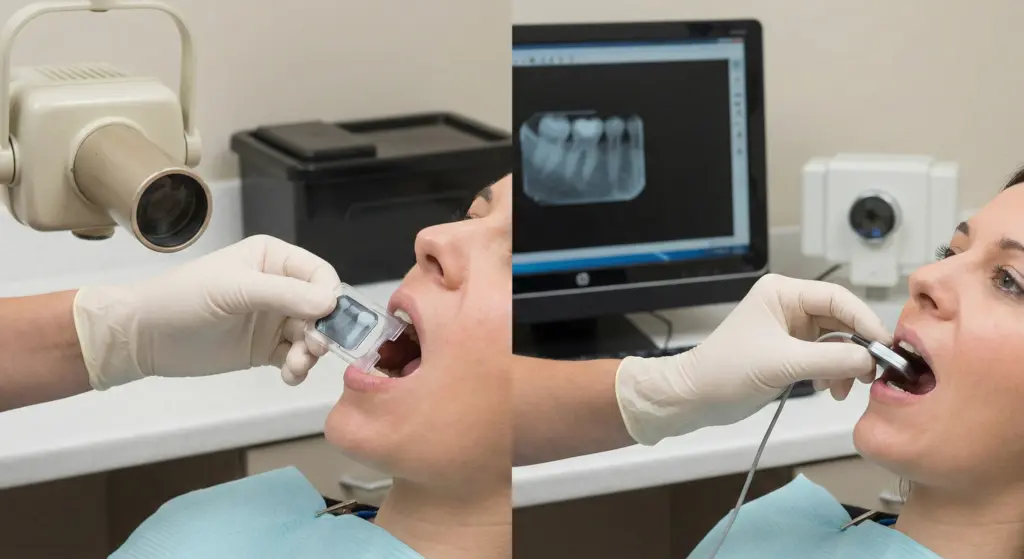
Digital Sensors vs. Film
Modern digital dental X-rays use electronic sensors. These sensors are far more sensitive, requiring significantly less radiation to capture high-quality images. Exposure times are much shorter often fractions of a second.Digital dental X-rays generally use significantly less radiation than older film X-rays, though the exact reduction depends on the equipment and settings.
Instant Results and Better Quality
Digital images appear on screens within seconds, eliminating re-takes due to processing errors and reducing unnecessary exposure. Dentists can enhance images, zoom on specific areas, and share easily for consultations.
Focused Beams and Collimation
Modern equipment uses collimation focusing X-ray beams only on specific areas being imaged. This prevents unnecessary exposure to surrounding tissues and further reduces overall doses.
How Dentists Keep Radiation Low: The ALARA Principle
Pediatric dentists follow ALARA: ‘As Low As Reasonably Achievable.’ This means using the lowest radiation dose while obtaining needed diagnostic information.
Risk-Based Imaging Decisions
X-rays aren’t automatic. Dentists consider:
- Age and developmental stage
- Cavity risk (diet, hygiene, previous decay, fluoride exposure)
- Symptoms (pain, swelling, trauma)
- What visual exams cannot reveal
- Time since last X-rays
Child-Specific Settings
Machines adjust for children’s smaller bodies, using lower exposure settings, fast exposure times, and precise positioning to capture needed areas without unnecessary exposure.
Trained Staff
Professionals receive specialized training in radiation safety, positioning techniques, and equipment operation. Proper positioning on the first try avoids retakes and minimizes total exposure.
Do Kids Need Lead Aprons or Thyroid Collars?
This common question has evolved with modern technology and updated professional guidance.
Updated Guidance on Shielding
With modern, collimated digital dental X-rays, routine patient shielding (lead aprons and thyroid collars) is not always necessary. Current recommendations emphasize minimizing exposure by optimizing technique using tight collimation, correct positioning, and child-specific settings while following the ALARA principle and taking X-rays only when clinically needed.
That said, practices vary some offices still use shielding for comfort or because of local/state rules. If you prefer a thyroid collar, ask your dentist what they use and why.
How Often Should Children Get Dental X-Rays?
Frequency depends on individual needs, risk factors, and dental health status.
General Guidelines
- Low cavity risk: Bitewing X-rays every 12-24 months
- Moderate to high risk: X-rays every 6-12 months to monitor decay
- New patients: Baseline X-rays unless recent ones are available
- Panoramic X-rays: as clinically indicated
Common Myths vs. Evidence-Based Facts
|
Myth
|
Evidence-Based Fact
|
|---|---|
|
Dental X-rays aren't safe for children.
|
Modern dental X-rays use very low radiation. Dentists recommend them only when diagnostic benefits outweigh minimal exposure.
|
|
Without lead aprons, it's unsafe.
|
Modern equipment with proper technique is safe with or without routine shielding. Many offices still use aprons, which is also fine.
|
|
X-rays are just for cavities.
|
X-rays assess development, eruption, infections, trauma, and orthodontic needs—issues not visible during visual exams.
|
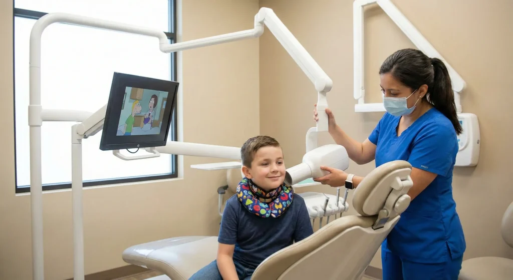
What Happens During a Child's Dental X-Ray
Understanding the process eases anxiety:
- Positioning: The assistant helps your child sit comfortably and positions a small sensor in their mouth
- Quick exposure: The machine is positioned and the X-ray taken in a fraction of a second, just a click
- Protection: Depending on practice, a lead apron may be used
- Review: Images appear on screen immediately; retakes are rare with trained staff
Pediatric teams use gentle language, demonstrate on parents, offer praise, and work quickly. If your child is anxious, let the team know ahead of time.
Frequently Asked Questions
Yes, when used appropriately. Pediatric dentists use child-specific settings with extremely low radiation doses.
Visual exams detect surface cavities but cannot see between teeth where they touch. Decay also occurs beneath surfaces or fillings.
It depends on cavity risk. Low-risk children may have bitewings every 12-24 months; higher-risk children every 6-12 months. Panoramic X-rays are taken as clinically indicated, often for growth/development assessment or orthodontic planning.
Yes, digital systems typically use significantly less radiation than older film X-rays Digital sensors are more sensitive, requiring shorter exposure times while providing high-quality images.
Yes, but inform your dentist about recent medical imaging. They can factor cumulative exposure into decisions, though dental X-rays use very low doses compared to most medical imaging.