
Gentle Pediatric Dentistry: What Makes a Kids’ Dental Visit Different
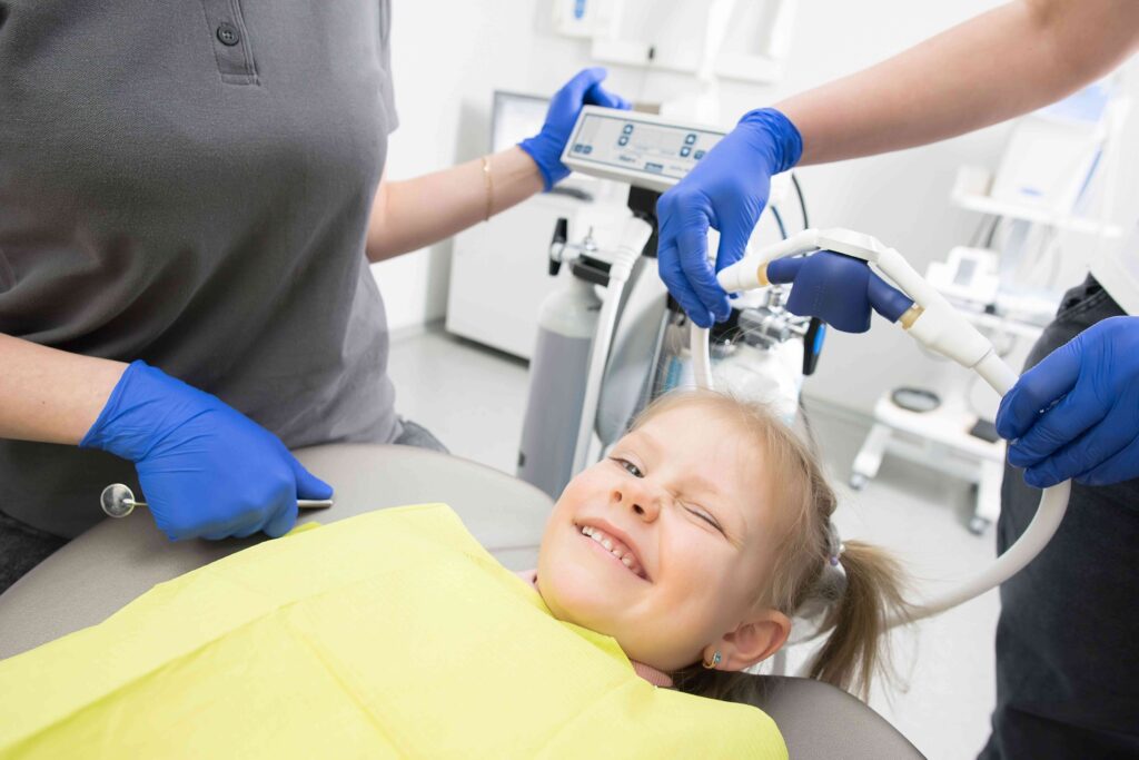
Gentle Pediatric Dentistry Guide · Williamsburg Brooklyn A child’s dental visit is not a smaller version of an adult appointment. It is designed around how children think, feel, and respond — with age-appropriate communication, careful pacing, and a focus on prevention and positive experience-building. Gentle Pediatric Dentistry: What Makes a Kids’ Dental Visit Different BOOK A PEDIATRIC VISIT FIRST VISIT GUIDE Written by Toothology Editorial Team | Reviewed by Dr. Ayala Lieberman, DDS September 2026 Some children have been to the dentist before and remember something that frightened them — a sound, a sensation, not knowing what was coming next. Others are going for the first time and do not know what to expect at all. Gentle pediatric dentistry is about more than speaking softly. It combines age-appropriate communication, careful pacing, individualized behaviour guidance, and a strong focus on prevention — adapting the visit to each child’s comfort level and previous experiences. What “Gentle” Actually Means in Pediatric Dentistry When parents look for a “gentle” dentist, they usually want someone who works at the child’s pace, explains treatment in age-appropriate language, and responds calmly to fear or resistance. In pediatric dentistry, this approach is supported by behaviour-guidance techniques such as tell-show-do, positive reinforcement, distraction, and desensitisation. “Gentle” does not guarantee that every visit will be completely comfortable. It means the dental team adapts its approach to the child’s age, temperament, previous experiences, treatment needs, and individual concerns. The goal is a predictable, confidence-building experience — not a rushed appointment. WHAT PARENTS ARE REALLY LOOKING FOR When parents search for a “gentle” kids’ dentist, they most often want someone who won’t push through resistance, will explain what’s happening, and will treat their child as an individual rather than a procedure to complete. These expectations are reasonable — and they describe the standard approach in thoughtful pediatric dental care. How a Kids’ Visit Differs From an Adult Visit A pediatric dental visit is designed around the developmental needs of a child, not just around the procedures involved. Several elements of the appointment work differently from an adult appointment. Communication Children are spoken to in age-appropriate terms — not clinical language, but not so simplified as to be condescending. The goal is making the child an active participant in their own care. Pacing Pediatric visits may build in extra time to allow a child to adjust to each step, ask questions, or take a short break. The dentist adapts to the child’s readiness rather than a fixed schedule. Prevention & Parent Education A significant portion of the visit focuses on preventing future problems. Because parents manage a young child’s oral health at home, guidance is directed at the family as much as the child. Building a Foundation A positive, well-explained visit can help children become more comfortable with dental care over time — creating the conditions for a lifetime of easier appointments. Aspect Adult Visit Kids’ Visit Communication Clinical language, direct and efficient Age-appropriate terms, tell-show-do, child as active participant Pacing Follows a set appointment schedule Builds in extra time; adapts to the child’s readiness at each step Primary Focus Treatment, restorations, and hygiene Prevention, parent education, and positive experience-building Behaviour Support Not typically part of the visit Communication techniques, desensitisation, distraction, nitrous oxide if appropriate Parent Involvement Usually minimal Parents included in home care guidance and oral health education 💡 The exact visit depends on the child’s age, comfort level, treatment needs, and previous dental experiences — no two pediatric visits are identical. Helping an Anxious or Previously Frightened Child Dental anxiety in children can come from a previous difficult visit, unfamiliar sounds or sensations, fear of needles, or simply not knowing what to expect. No single approach works for every child — but several techniques consistently help. Share concerns before the visit. Tell the dental team about previous negative experiences, sensory sensitivities, or specific triggers. This allows the team to prepare and adapt before the child arrives. Use positive, neutral language. Keep explanations calm and simple. Avoid promising “it won’t hurt” — this can backfire. Instead: “The dentist will explain what is happening and take good care of you.” Tell-show-do. The dentist explains a step, may demonstrate what to expect, and then proceeds. This removes the element of surprise that often amplifies anxiety. Gradual desensitisation. For very anxious children, a slower introduction to the dental environment — getting familiar before treatment begins — can make future visits significantly easier. Comfort items and distraction. A familiar toy or object, headphones, or a video can help a child manage their attention during treatment. BEHAVIOUR GUIDANCE TECHNIQUE TELL — SHOW — DO STEP 1 TELL The dentist explains what will happen in simple, reassuring language appropriate to the child’s age. STEP 2 SHOW The dentist demonstrates the instrument or step — letting the child see, hear, or feel it before proceeding. STEP 3 DO Only then does the dentist proceed with the step — giving the child a sense of control and predictability. What Happens at a First Dental Visit The ADA and AAPD recommend establishing dental care after the first tooth appears and no later than the first birthday. Early visits help establish a dental home, assess oral health, guide parents on hygiene and diet, and introduce the child to the dental environment in a low-pressure way. Review of health and dental history Meeting the dental team and becoming familiar with the office An age-appropriate oral examination Cavity-risk discussion and oral health assessment Brushing technique and diet guidance for parents Cleaning when appropriate for the child’s age and comfort Fluoride when clinically appropriate X-rays only when clinically indicated GOAL: PREDICTABLE AND POSITIVE The goal of a first visit is to make the experience feel predictable and positive — not to rush through a full set of procedures. What happens at the appointment depends on the child’s age, comfort level, any symptoms present, and their developmental readiness on the day. When Nitrous Oxide May Be Considered Nitrous oxide — sometimes called laughing gas — may help
Dental Crown Pain After Root Canal: Is It Normal or a Warning Sign?

Crown & Root Canal Clinical Guide · Brooklyn Root canal treatment is complete and the crown is in place — but the tooth still hurts. Mild tenderness can be part of normal healing. The key question is whether your discomfort is improving or getting worse. Dental Crown Pain After Root Canal: Is It Normal or a Warning Sign? BOOK AN EVALUATION SEE THE URGENCY GUIDE Written by Toothology Editorial Team | Reviewed by Dr. Ayala Lieberman, DDS September 2026 Dental crown pain after root canal treatment does not automatically mean the treatment has failed. Mild tenderness can occur while the tissues around the tooth heal. What matters most is the pattern — whether discomfort is steadily improving or becoming more persistent and intense. Mild and Improving Often consistent with normal post-treatment healing. Monitor and follow aftercare instructions. Lasting More Than a Few Days Should be evaluated. Pain that doesn’t resolve on its own within the expected window needs a dentist’s review. Pain That Returns After Improving A warning sign. Pain coming back after a symptom-free period should be assessed promptly. Severe or With Swelling Should not be ignored. Severe pain or any swelling alongside crown discomfort needs prompt dental evaluation. Is Crown Pain After a Root Canal Normal? Mild soreness for a few days after root canal treatment can be normal, especially if the tooth was painful or infected beforehand. This tenderness usually comes from the tissues around the root — not the tooth itself — and should gradually improve as healing progresses. Contact your dentist if the pain is any of the following: Severe or worsening over time Lasting more than a few days without improving Returning after the tooth had felt better Accompanied by swelling, pus, or bad taste Triggered by an uneven or high bite THE PATTERN MATTERS MORE THAN THE PAIN LEVEL A tooth that is mildly sore but improving each day is very different from one that is worsening, plateauing, or getting better then worse again. Tracking the direction of your symptoms is the most useful thing you can do before your follow-up appointment. Why a Treated Tooth Can Still Hurt A root-canal-treated tooth can still hurt because the tissues around the root remain sensitive, even though the nerve tissue inside the tooth has been removed. Several distinct issues can cause pain under a crown, and many of them feel similar — which is why a dental exam is usually needed to identify the actual source. Normal healing: Temporary inflammation around the root tip after treatment — should improve steadily over days. High bite: A crown that contacts the opposing tooth too early or too heavily places extra pressure on the tissues around the root, causing soreness when biting. Crown fit or cement problems: A loose or poorly sealed crown can allow bacteria to reach the underlying tooth structure. Inflammation at the root tip: Persistent inflammation around the apex can cause ongoing tenderness or pressure sensitivity. Crack or fracture: A damaged tooth or root may cause pain specifically when chewing or applying pressure. Reinfection or new decay: Bacteria entering through decay or a leaking crown can cause pain months or even years after the original treatment. Crown Pain When Biting or Chewing Some sensitivity when biting is expected during early healing after a root canal and should gradually improve. If the crown hurts when biting and that pain persists, worsens, or starts after the tooth had felt better, possible causes include a high bite, a crack or fracture, ongoing inflammation or reinfection, or a loose or leaking crown. Because these problems feel similar, bite pain alone cannot confirm the cause. If discomfort is not steadily improving, schedule a dental evaluation rather than continuing to test the tooth by chewing on it. DO NOT FILE OR GRIND THE CROWN AT HOME Even if the crown feels like it might be too high, do not try to adjust it yourself. Bite adjustments need to be done by a dentist using articulating paper so that only the necessary contact points are changed — not the crown’s overall shape or seal. Could the Bite Be Too High? A high crown contacts the opposing tooth too early or too heavily. This extra pressure irritates the tissues around the root and causes soreness — especially when biting. The effect can be cumulative: repeated pressure on already-healing tissues slows recovery and makes symptoms feel more pronounced. A bite problem is not always obvious. Even when the crown does not feel dramatically “off,” it may still be receiving more pressure than it should. The dentist can check the bite with articulating paper and make a small, precise adjustment if needed. This is one of the most common and most easily corrected causes of post-treatment soreness. Can a Root Canal Tooth Become Reinfected? Yes. A root-canal-treated tooth can become reinfected — sometimes months or years after treatment seemed successful. Pain that returns after a symptom-free period should be evaluated promptly. Possible causes of reinfection include: Missed canal anatomy: Complex root systems may include canals that were not fully treated initially Delayed or failed seal: Bacteria may re-enter if the tooth was not permanently restored soon enough after the root canal New decay: Cavities around crown margins can allow bacteria to reach the canal system Loose or damaged restoration: A cracked crown or filling can compromise the internal seal Tooth fracture: A crack extending into the root may allow persistent bacterial contamination RETREATMENT MAY BE POSSIBLE If reinfection is confirmed, endodontic retreatment — reopening, cleaning, and resealing the canals — may be an option. Whether it is appropriate depends on the tooth structure, extent of infection, existing restoration, and whether a fracture is present. A dental exam and imaging are needed to assess suitability. When Crown Pain Becomes an Emergency Not every symptom requires emergency care. Use the pattern below to decide when to monitor at home, contact the dentist, or seek immediate medical attention. Monitor / Improving ✔ Call The Dentist ☎ Emergency Care 🚨
Temporary Dental Crown Fell Out: What to Do Before Seeing a Dentist

Emergency Dental Guide · Williamsburg Brooklyn If your temporary or permanent crown fell out, do not panic — but do act quickly. Save the crown, protect the tooth, and call the dentist. The steps you take in the next hour matter. Dental Crown Fell Out: What to Do Before Seeing a Dentist CALL 718-678-3800 WHAT TO DO FIRST Written by Toothology Editorial Team | Reviewed by Dr. Ayala Lieberman, DDS September 2026 If your temporary dental crown fell out, do not panic — but contact your dentist as soon as possible. Whether it is a temporary or permanent crown, the tooth underneath needs protection and should be checked promptly. The most important thing you can do right now is save the crown, avoid chewing on that side, and make the call. Dental Bridge Symptoms — Quick Reference Do First Save the crown in a clean container, rinse gently, avoid chewing on that side, and call the dentist Do Not Use super glue, force the crown back on, or eat sticky or hard foods on that side Urgent Signs Severe pain, swelling, fever, trauma, bleeding, or signs of infection Best Next Step Book an emergency or restorative dental evaluation at Toothology in Williamsburg Why Temporary Crowns Come Loose Temporary crowns are held with temporary dental cement, which is intentionally weaker than permanent cement — because the temporary crown must be removable when the final crown is ready. A temporary or permanent crown may come loose for several reasons: Sticky or chewy foods— gum, caramel, or taffy can pull the crown off Hard foods— biting down hard can shift or loosen the crown Grinding or clenching— extra pressure can weaken the cement or crack the crown over time Flossing technique— pulling floss straight upward can lift a temporary crown; slide it out sideways instead Cement breakdown— temporary cement weakens if the crown has been in place for several weeks Decay or fracture— changes to the tooth structure underneath can affect the fit and cause the crown to loosen IT IS NOT ALWAYS YOUR FAULT A crown coming loose is not always the patient’s fault. Temporary cement has limits by design, and even permanent crowns can come off over time with normal wear. The important thing is to protect the tooth and contact your dentist promptly. What to Do Right Away If a Crown Falls Out These steps apply whether a temporary crown or a permanent crown came off: Find and save the crown. Place it in a clean container or small bag and bring it to your appointment — even if a new crown may be needed. Rinse gently. Use warm water to clear debris. Avoid aggressive rinsing if the tooth is sensitive or you have bleeding. Check it, but do not force it. If the crown looks cracked, badly dirty, or does not sit easily over the tooth, leave it out and bring it to the dentist. Avoid chewing on that side. Eat soft foods and use the opposite side until the dentist checks the tooth. Call the dentist promptly. Tell them whether it was a temporary or permanent crown, whether you have the crown, and whether you have pain, swelling, sensitivity, or a sharp edge. Bring the crown with you. Even if a new crown is needed, the old one helps the dentist assess what happened and whether the fit has changed. What Not to Do With a Fallen Crown Do not use super glue, nail glue, craft glue, or any household adhesive — they are unsafe in the mouth and can damage the tooth or crown permanently Do not force the crown back on — if it does not fit easily and comfortably, leave it out and bring it to the dentist Do not chew on the exposed tooth or on the side where the crown is missing Do not use sharp tools, toothpicks, or metal objects to probe or clean the exposed tooth Do not place aspirin or tablets directly on the tooth or gum tissue Do not ignore severe pain, swelling, fever, bad taste, or any sign of infection Do not assume everything is fine because there is no pain — an exposed tooth is still vulnerable to sensitivity, bacteria, and fracture Can You Put the Crown Back on Yourself? The safest answer is to call your dentist and get the crown checked promptly. A crown should only be recemented after the dentist confirms that both the tooth and the crown still fit properly. Do not assume that because the crown feels like it fits that it is safe to self-cement permanently. In some situations, an over-the-counter temporary dental cement may be used as a very short-term measure until your appointment. This is not a permanent fix. Only consider it if all of the following are true: The crown sits easily over the tooth without forcing or pressure The crown is not cracked, chipped, or badly discoloured The tooth does not hurt when the crown is placed over it You are using a product specifically made for temporary dental use — and following the instructions exactly You can still contact a dentist for proper evaluation as soon as possible TEMPORARY CEMENT IS NOT A FIX Even if temporary cement makes the crown feel stable, the tooth still needs a professional evaluation. The dentist must confirm the fit, check for decay or fracture, and determine whether the same crown can be used again or whether a new one is needed. Is a Fallen Crown a Dental Emergency? A crown coming off should always be reported to the dentist promptly, but the urgency depends on what you are experiencing. Use the guide below to assess how quickly to act. Situation What It May Mean How Urgent? 🪥 Crown Off, No Pain, No Damage Cement failed or temporary cement reached its limit — common and manageable Call today — same-day or next-day appointment 🌡 Tooth Sensitive to Air or Temperature Exposed dentine or nerve — the tooth is unprotected and needs coverage Call today
Dental Bridge Infection Symptoms: When Pain, Swelling, or Bad Taste Needs Attention

Restorative & Emergency Dental Guide · Brooklyn Pain under a bridge, swelling near the gumline, a bad taste, or looseness should not be ignored. These symptoms may point to a problem around the supporting tooth, gum tissue, or cement margin — and early evaluation almost always leads to simpler treatment. Dental Bridge Infection Symptoms: When Pain, Swelling, or Bad Taste Needs Attention CALL 718-678-3800 SEE URGENCY GUIDE Written by Toothology Editorial Team | Reviewed by Dr. Ayala Lieberman, DDS September 2026 Pain under a dental bridge, swelling near the gumline, a bad taste, odor, or looseness should not be ignored. These symptoms may point to a problem around the supporting tooth, gum tissue, or cement margin beneath the bridge. Not every symptom means a severe emergency, but dental bridge problems should be checked promptly — an exam and X-rays can identify whether the issue is decay, gum inflammation, infection, a loose bridge, or another concern. Dental Bridge Symptoms — Quick Reference Signs to Evaluate Pain, swelling, bad taste, odor, bleeding gums, looseness, or pain when chewing Urgent Symptoms Severe pain, fever, pus with swelling, facial or jaw swelling, or a loose or fallen bridge Emergency Medical Care Trouble breathing or swallowing, or rapidly spreading facial or neck swelling Do Not Try to remove the bridge, use household glue, ignore swelling, or keep chewing on a painful or loose bridge How to Tell If a Dental Bridge Is Infected The phrase “dental bridge infection” can be misleading. The bridge itself does not become infected. The problem usually develops around the supporting teeth, gum tissue, bone, or the space beneath the artificial tooth — and symptoms can start mildly before getting worse. Possible signs that something is wrong around a dental bridge: Persistent or throbbing pain— especially near one side of the bridge Swelling— puffiness or tenderness near the gumline or bridge margin Pus or discharge— a gum bump or drainage with an unpleasant taste or odor Bad taste or odor— bacteria, decay, or drainage under the bridge may cause a persistent foul taste or smell Bleeding gums— gum inflammation around the bridge or supporting teeth Sensitivity— unusual pain with temperature changes, pressure, or chewing SYMPTOMS CAN DEVELOP SLOWLY Decay or infection under a bridge may not always cause obvious pain right away. Regular dental exams with X-rays help detect problems while they are still easier to treat — even when symptoms are absent or mild. Pain Under a Bridge: What It Usually Means Tooth pain under a bridge can come from several different causes — not always infection. Because the symptoms can feel similar, dental bridge pain should be checked with an exam and X-rays rather than relying on pain relievers alone. Decay under the bridge: Cavities can form around the supporting teeth or bridge margins, especially in areas difficult to clean. Loose cement or margin failure: A broken seal lets bacteria enter under the bridge. Gum inflammation: Irritated or infected gum tissue can cause pain, bleeding, or sensitivity around the bridge. Cracked abutment tooth: A tooth supporting the bridge may crack under bite pressure — sometimes without obvious visible damage. Root infection or abscess: Deep decay or infection reaching the root can cause pain in the area beneath the bridge. Bite misalignment: A high or uneven bite can cause chewing pain even without decay or infection. Swelling, Odor, and Bad Taste as Warning Signs Swelling near a dental bridge, a persistent bad taste, or an odor that does not improve with brushing should be evaluated by a dentist. Swelling may indicate gum infection, a periodontal abscess around a supporting tooth, or a dental abscess at the tooth root. Bad taste or odor can result from trapped food, bacterial buildup under the bridge, decay, or drainage from an infected area. A bad taste alone does not necessarily mean there is a serious infection. However, if it occurs alongside swelling, pain, pus, tenderness, or a painful bump near the bridge, contact your dentist promptly. When to Seek Emergency Medical Care If swelling spreads rapidly toward the face or neck, or if you have difficulty breathing or swallowing, seek emergency medical care immediately — not a dental appointment. These can be signs of a spreading dental infection that requires urgent treatment. Why Decay Can Form Beneath a Bridge The teeth supporting a dental bridge can still develop cavities, especially around areas that are harder to clean. Plaque commonly builds up at the crown margins near the gumline, beneath the pontic, and between the bridge and neighboring teeth. Because a regular toothbrush cannot fully clean under a fixed bridge, additional tools may help: Floss threaders or superfloss — threaded under the bridge for contact point cleaning Water flossers — useful for flushing debris from beneath the pontic and around margins Small interdental brushes — for cleaning around crown margins near the gumline ASK YOUR HYGIENIST A dentist or hygienist can show you the best cleaning technique for your specific bridge design at your next visit. Without consistent cleaning, plaque can gradually damage the supporting teeth and cavities may develop before noticeable symptoms appear. When a Bridge Becomes Loose A loose dental bridge should be evaluated promptly. Common causes include cement failure, decay in a supporting tooth, fracture, excessive bite pressure, or normal wear over time. If the bridge feels loose, moves, or comes out: IF YOUR BRIDGE IS LOOSE — DO THIS Avoid chewing on that side to reduce the risk of further damage Save the bridge or any loose pieces in a clean container if they come out Rinse gently with warm water Contact your dentist promptly — especially if you also have pain, swelling, bleeding, or sharp edges DO NOT DO THIS Do not use super glue or household adhesives to reattach the bridge Do not pull on a partially attached bridge Do not keep chewing on the bridge or affected side Do not wait if you have pain, swelling, or the bridge has fully come out
White Spots on Teeth After Braces: Can a Cosmetic Dentist Fix Them?
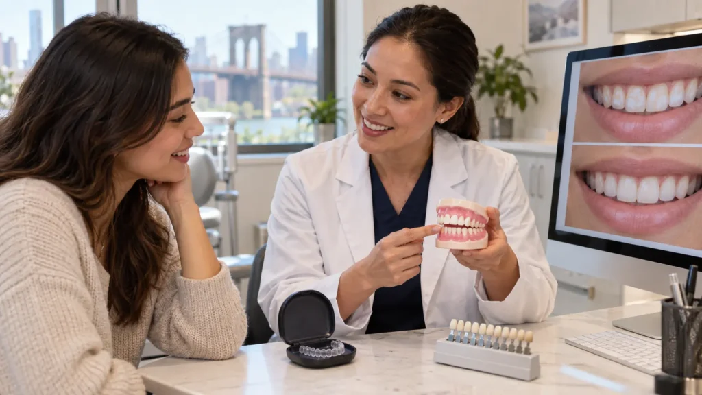
Post-Braces Cosmetic Guide · Williamsburg Brooklyn Chalky white patches where your brackets used to be are common after braces — and a cosmetic dentist may be able to help. The right treatment depends on the cause, depth, and whether active enamel changes are present. White Spots on Teeth After Braces: Can a Cosmetic Dentist Fix Them? BOOK A POST-BRACES CONSULT SEE TREATMENT OPTIONS Written by Toothology Editorial Team | Reviewed by Dr. Ayala Lieberman, DDS September 2026 After braces come off, some patients notice chalky white patches where the brackets used to be. These white spots on teeth after braces are a common post-orthodontic concern — and they can affect how the final smile looks after all that work. A cosmetic dentist may be able to improve them, but the right treatment depends on the cause, depth, and whether active enamel changes are present. The best first step is a post-braces dental evaluation. White Spots After Braces at a Glance Common Cause Enamel demineralization around brackets or hard-to-clean areas — where plaque and acids weakened the tooth surface during treatment. Not Always Permanent Mild surface spots may fade over time. Deeper or more established lesions often need professional treatment to improve. Whitening Is Usually Not the Fix Whitening changes the overall tooth shade but does not repair enamel mineral loss — and may temporarily make spots more noticeable. Possible Treatments Fluoride, remineralization support, microabrasion, resin infiltration, or dental bonding — depending on cause, depth, and activity of the spots. Why White Spots Appear After Braces Most white spots after braces are caused by enamel demineralization. This happens when plaque bacteria produce acids that pull minerals from the tooth surface, leaving enamel weak, chalky, or opaque in appearance. Brackets and attachments can make certain areas harder to clean. If plaque stays around the bracket edges or near the gumline for extended periods, white spot lesions can develop in those areas. Braces themselves do not directly cause the spots — the issue is plaque and acid exposure around the appliances, which is why some patients finish orthodontic treatment without any spots while others notice visible changes. White spots most commonly appear around bracket edges, along the gumline, or on the front teeth where they are most visible. Are White Spots After Braces Early Cavities? They can be, but not always. White spots often represent early enamel demineralization — where minerals have been lost from the tooth surface but a cavity has not yet fully formed. This stage is sometimes described as a “pre-cavity.” With the right care, some early white spot lesions may be stabilised or improved. Ignored, some can progress into actual cavities over time. NOT EVERY WHITE PATCH IS FROM BRACES Some white spots have nothing to do with orthodontic treatment. A post-braces dental exam helps determine whether the spots are active demineralization, cosmetic discoloration, a pre-existing enamel condition, or early decay that needs restorative care. Other Possible Causes of White Spots FLUOROSIS White streaks or patches that developed while the teeth were forming due to fluoride exposure. These are pre-existing and unrelated to braces. ENAMEL HYPOPLASIA Areas where enamel formed thin, weak, or uneven during tooth development — often present before braces and more visible after treatment. OLD TRAUMA A previous tooth injury that caused localised enamel changes — which may become more noticeable after teeth are straightened and more visible. Will White Spots Fade on Their Own? Mild, very superficial white spots may become less noticeable over time — especially once brushing becomes easier after brackets are removed. Better oral hygiene, enamel rehydration, fluoride, and remineralization support may help very surface-level spots blend in more naturally. However, many established white spot lesions do not fully disappear on their own. The enamel may become stronger, but the chalky or opaque appearance can remain. For mild spots, monitoring over a few months with excellent home care is reasonable. If spots are deeper, very noticeable, or unchanged after consistent oral hygiene, a professional dental evaluation is the right next step. Why Whitening Usually Does Not Fix White Spots Whitening is often the first thing patients think of — but it is usually not the best starting point for white spots after braces. Whitening lightens the overall tooth shade. Since white spot lesions are already lighter than the surrounding enamel, whitening may temporarily make the contrast more noticeable rather than reducing it. Whitening also does not repair enamel mineral loss, stop active demineralization, or close an early cavity. For this reason, white spots should be evaluated before starting any whitening treatment. A dentist can confirm whether the spots are cosmetic, active demineralization, or early decay — and then recommend the right sequence of care. EVALUATE BEFORE YOU WHITEN Whitening may be appropriate as part of a broader cosmetic plan — but after the white spots are properly assessed and treated, not before. Starting with a dental evaluation protects the enamel and leads to a better cosmetic result. Remineralization and Minimally Invasive Options For many white spots after braces, the first goal is to strengthen and stabilise the enamel before choosing a cosmetic treatment. The right option depends on the depth, size, activity, and location of the spots. Fluoride Varnish Professionally applied fluoride may support remineralisation and help protect enamel from further acid damage. Often recommended as a first-line supportive step for active or significant spots. Prescription Fluoride Toothpaste A dentist may recommend stronger fluoride toothpaste for active or significant demineralisation, used at home as part of a daily preventive routine. Calcium-Phosphate Remineralisation Support Products such as MI Paste may support remineralisation in selected cases. These should only be used if specifically recommended by the dentist — not self-prescribed. Microabrasion A minimally invasive option that removes a very thin outer enamel layer to improve shallow surface discoloration. Suitable only for very superficial spots. Resin Infiltration A treatment that may help mask some non-cavitated white spot lesions with minimal tooth preparation. Confirm availability and suitability at your consultation — not every spot qualifies. ACTIVE
Dental Onlay vs Crown: Which Is Better for a Damaged Tooth?
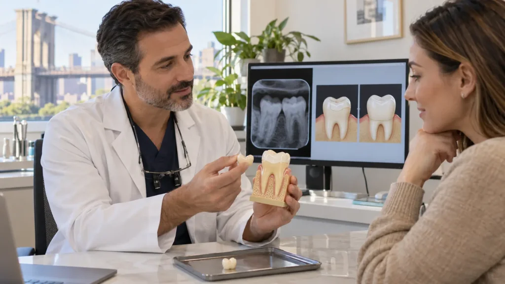
Restorative Dentistry Guide · Williamsburg Brooklyn If a filling is no longer enough, a crown isn’t automatically the next step. A dental onlay may preserve more tooth structure when the conditions are right. The difference comes down to what the tooth can actually support. Dental Onlay vs Crown: Which Is Better for a Damaged Tooth? SEE THE COMPARISON WHEN EACH IS USED Written by Toothology Editorial Team | Reviewed by Dr. Ayala Lieberman, DDS September 2026 If your dentist says a filling is no longer enough, you may be wondering whether a crown is your only option. This is where the onlay vs crown comparison matters. A dental onlay can be a more conservative choice when the tooth still has enough healthy structure. A dental crown may be better when the tooth is badly weakened, deeply decayed, or heavily cracked. Neither is automatically better — the right choice depends on the tooth. Onlay vs Crown: At a Glance Dental Onlay Covers part of the tooth and one or more cusps. May preserve more healthy tooth structure when the tooth is suitable for a partial restoration. Dental Crown Covers the full visible portion of the tooth from the gumline up. Provides stronger protection when the tooth is severely damaged or weakened. Neither Is Always Better The right option depends on the condition of the tooth — not a preference or a cost choice. A dental exam determines what the tooth can safely support. How the Decision Is Made Exam findings and X-rays guide the recommendation — the amount of remaining tooth structure, any crack pattern, bite forces, and the size of the damage all matter. What Is a Dental Onlay? A dental onlay is a custom restoration used to repair a damaged part of a back tooth. Unlike a standard filling placed directly in the mouth, an onlay is fabricated outside the mouth — either in a dental lab or with digital milling technology — and then precisely bonded to the prepared tooth. An onlay covers one or more cusps — the raised points of the tooth that handle biting and chewing force. Because of this, it is sometimes called a partial crown. It sits between a large filling and a full dental crown in terms of coverage and preparation. Onlays are commonly made from ceramic or porcelain for a natural appearance and good durability, though other materials may also be used depending on the case. Each onlay is custom-made to fit the prepared tooth, support the bite, and protect the damaged area. FILLING Repairs within the chewing surface only. No cusp coverage. ONLAY Covers chewing surface and one or more cusps. Partial coverage. CROWN Covers the full visible tooth from the gumline up. Full coverage. Onlay vs Crown: The Core Difference The main difference between a dental onlay and a dental crown is how much of the tooth each restoration covers — and how much natural tooth structure is removed to place it. Factor Dental Onlay Dental Crown Coverage Covers the damaged chewing surface and one or more cusps — partial coverage Covers the full visible portion of the tooth from the gumline up Tooth Preparation Preserves more natural tooth structure when the tooth is suitable Requires reshaping the tooth on all sides for full coverage Best For Moderate damage, large filling replacement, or fractured cusp when enough healthy tooth remains Severe damage, extensive decay, little structure left, root canal-treated teeth, or heavy bite forces Material Often ceramic or porcelain — matched to tooth colour Porcelain, zirconia, ceramic, or metal-based options depending on the case Process Exam, preparation, scan or impression, temporary if needed, and final bonded placement Similar process with full crown preparation, temporary, and final cementation Longevity Can last many years with good care and regular monitoring at dental visits Can also last many years — longevity depends on care, material, and bite forces IN SIMPLE TERMS An onlay is more conservative when the tooth can support it. A crown is more protective when the tooth needs full coverage. The exam — not a preference — determines which option is appropriate for that specific tooth. How Much Tooth Structure Each Option Removes Preserving natural tooth structure matters because enamel and dentin do not regenerate once they are removed. For that reason, dentists try to use the most conservative restoration that can safely repair the tooth — not the most conservative option regardless of suitability. SUITABILITY DETERMINES WHICH IS CONSERVATIVE An onlay only qualifies as the conservative option when the tooth can safely support it. If a tooth needs full coverage to remain functional and protected, a crown is the safer and more appropriate choice — placing a partial restoration on an unsuitable tooth is not conservative, it is under-treating the problem. When an Onlay May Be the Better Choice A dental onlay may be a good option when the tooth is too damaged for a filling but still has enough healthy structure to support a partial restoration safely. Created with Sketch Beta. Replacing a large old filling: If an old filling is cracked, worn, or has decay around its margins, an onlay may provide stronger support when enough healthy tooth structure remains. Fractured cusp: If one cusp is damaged but the rest of the tooth is stable and solid, an onlay can cover and protect that specific area. Moderate decay: If decay is too large for a filling but not extensive enough to require full crown coverage, an onlay may be a suitable middle option. Tooth structure preservation: When the tooth is clinically suitable, an onlay can restore strength and function while preserving more natural tooth than a crown would require. ONLAYS ARE OFTEN USED ON PREMOLARS AND MOLARS These back teeth handle heavy chewing forces and are common sites for large fillings or cusp fractures. A restorative dental exam confirms whether the remaining tooth is solid enough to support an onlay or whether a crown is the safer option. When a Crown Is Necessary Instead A dental crown may
Vaping After Dental Implant Surgery: Healing Risks and Recovery Timeline

Dental Implant Aftercare Guide · Williamsburg Brooklyn Can you vape after tooth extraction or dental implant surgery? The safest answer during early healing is no. Here’s why it matters, what the recovery timeline looks like, and what to watch for. Vaping After Dental Implant Surgery: Healing Risks & Recovery Timeline SEE RECOVERY TIMELINE Warning Signs to Watch Written by Toothology Editorial Team | Reviewed by Dr. Ayala Lieberman, DDS August 2026 Can you vape after tooth extraction or dental implant surgery? The safest answer is no, not during early healing. Vaping can interfere with blood clot formation, gum tissue healing, and the recovery process after oral surgery. When it is safe to resume depends on the procedure, whether bone grafting was involved, how healing is progressing, and your dental team’s specific instructions — not a general timeline you find online. IF YOU ALREADY VAPED AFTER SURGERY Call your dental office and explain what happened. The team can check the healing site, look for early signs of complications, and guide you on next steps. Do not wait and hope for the best — early evaluation is usually easier to manage than a delayed problem. Why Vaping Is a Concern After Oral Surgery Oral surgery healing depends on a stable blood clot, healthy gum tissue repair, and — for dental implants — gradual bonding between the implant and jawbone. Vaping can interfere with this process in several specific ways. Suction Drawing on a vape creates pressure in the mouth that may disturb the healing blood clot — the same reason straws are also restricted after oral surgery. Nicotine & Blood Flow Nicotine can narrow blood vessels and reduce circulation to healing gum and bone tissue — the same tissue that depends on good blood flow to repair itself. Heat & Aerosol Irritation Vape aerosol and heat may irritate soft tissues that are already in a sensitive state during the early recovery period after surgery. Dry Mouth Vaping may reduce saliva flow, which plays a role in supporting a healthier oral healing environment after surgery. How Nicotine Affects Healing and Osseointegration Nicotine can affect oral surgery healing by reducing blood flow and oxygen delivery to the tissues that need to repair. This applies to gum tissue after extraction, bone tissue after grafting, and the implant site itself. WHAT IS OSSEOINTEGRATION? Osseointegration is the process where a dental implant gradually bonds with the surrounding jawbone after placement — creating a stable foundation for the final crown or bridge. This process typically takes several months and depends on good blood supply, healthy bone, and minimal disruption. Anything that reduces circulation or increases tissue irritation during this period can raise the risk of complications. Nicotine may interfere with this healing process in several ways: Reducing blood flow to healing tissue and bone Slowing gum and bone repair after surgery Increasing irritation around the surgical site Affecting the body’s normal healing and immune response Raising the risk of complications during the osseointegration period VAPING IS STILL BEING STUDIED — BUT THE CONCERNS APPLY NOW Smoking has been linked with higher dental implant complication and failure risks. Vaping is still being studied independently, but because most vape products contain nicotine, the same healing concerns apply. Avoid vaping during the implant recovery period unless your dentist or oral surgeon specifically says it is safe to resume. Vaping vs Smoking: Is One Safer After Surgery? Vaping is not a safe alternative to smoking after tooth extraction or dental implant surgery. Removing cigarette combustion from the equation does not remove the main healing concerns for oral surgery recovery. VAPING AFTER SURGERY Nicotine exposure reduces blood flow to healing tissue Suction can disturb the healing blood clot Aerosol may irritate sensitive post-surgical gum tissue Dry mouth may affect the healing environment Not a safe alternative to smoking during recovery SMOKING AFTER SURGERY Well-documented links to higher implant complication risks Nicotine and combustion chemicals reduce circulation Suction equally disturbs healing blood clot Light smoking is not risk-free — nicotine amount still matters Both quantity and timing during recovery affect outcome Recommended Recovery Timeline There is no single safe date for vaping after tooth extraction or dental implant surgery. Use the timeline below as a general healing guide — not as clearance to resume vaping. Always follow your dental team’s specific instructions and ask before restarting. Recovery Stage What Is Happening Vaping Guidance First 24 Hours Blood clot forms and tissue begins sealing — the most critical phase for clot stability Avoid vaping, smoking, straws, spitting, and any suction Days 2–3 Swelling and discomfort may peak; dry socket risk remains elevated Continue avoiding vaping — call the dentist immediately if pain is worsening First Week Soft tissue begins closing over the site; follow-up visit may be scheduled Ask your dentist at your follow-up before considering any resumption Weeks 2–6 Gum tissue matures; bone and implant healing continue beneath the surface Nicotine may still slow healing — especially after grafting or implant placement 3–6 Months Osseointegration continues as the implant gradually bonds with the surrounding jawbone Discuss vaping, smoking, and long-term implant risk with your dental team Use this timeline as a general healing guide only — not as clearance to resume vaping. Always follow your dental team’s specific aftercare instructions and ask before restarting. SURFACE HEALING DOESN’T MEAN COMPLETE HEALING Even if the extraction site or gum tissue looks healed from the outside, the deeper bone and tissue underneath may still be recovering — especially after implant placement or bone grafting, which can take several months. Always get clearance from your dental team before vaping again. Signs of Delayed Healing or Dry Socket Dry socket occurs when the blood clot in an extraction site is disturbed or fails to form properly — exposing the bone and nerves underneath and causing significant pain. Healing problems after implant placement should also not be managed at home. Contact your dental office promptly if you notice any of these: Pain that gets worse instead of gradually improving after the first
Dental X-Rays in Brooklyn: How Often Do You Really Need Them?
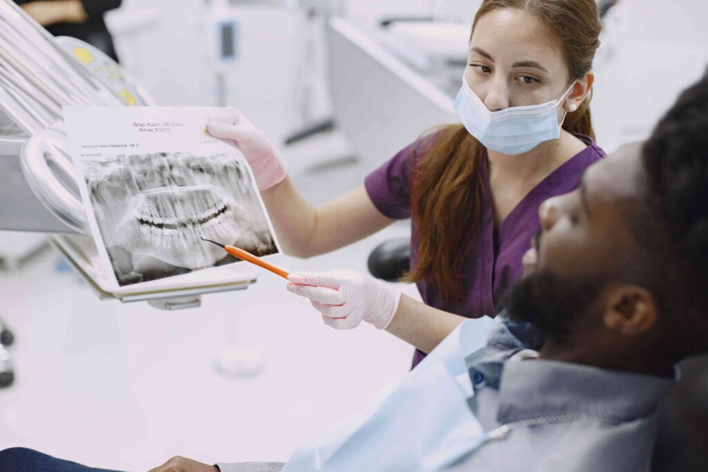
Patient Education Guide · Williamsburg Brooklyn No universal schedule applies to every patient. How often dental X-rays are recommended depends on your oral health, cavity risk, symptoms, and whether recent images are already available. Dental X-Rays in Brooklyn: How Often Do You Really Need Them? X-RAYS AT A GLANCE HOW OFTEN BY SITUATION Written by Toothology Editorial Team | Reviewed by Dr. Ayala Lieberman, DDS August 2026 Do you really need dental X-rays at every visit? Are they safe? These are reasonable questions. There is no single schedule that applies to every patient. How often dental X-rays are recommended depends on your age, oral health history, cavity risk, gum health, symptoms, and whether recent usable images are already available. X-rays help dentists see what a visual exam cannot always show — and they are used to support diagnosis, not as an automatic step at every appointment. Dental X-Rays at a Glance How Often Depends on oral health, symptoms, cavity risk, history, and whether recent usable images are already available — not a fixed schedule. Why Dentists Take Them To check for cavities between teeth, bone loss, infections, impacted teeth, and hidden issues a visual exam cannot fully see. Safety Dental X-rays use low radiation and should be recommended when they provide useful diagnostic information — following the ALARA principle. Digital X-Rays Faster, easier to store and share, and often lower in radiation than older film methods. Toothology uses digital imaging for all X-rays. BRINGING RECENT X-RAYS FROM ANOTHER OFFICE If you are visiting Toothology for the first time, bring or send any recent dental X-rays from your previous dental office. If they are current and clear, they may reduce or eliminate the need for new images at your first visit. Why Dentists Take Dental X-Rays A dental exam and a dental X-ray work together, but they do different things. During the exam, the dentist checks what can be seen directly — your gums, visible tooth surfaces, bite, soft tissue, and jaw function. X-rays show what cannot be fully seen from a visual exam alone. Cavity Detection Finding small cavities between teeth before they become larger — earlier detection usually means simpler, more conservative treatment. Bone Level Monitoring Checking for bone loss around tooth roots related to gum disease — bone changes are invisible to a visual exam. Tooth Infections Identifying infection or abscesses near the tooth root — often present without obvious visible signs. Development & Eruption Checking how children’s permanent teeth or wisdom teeth are developing and whether intervention may be needed. Treatment Planning Supporting care such as extractions, root canals, implants, or orthodontic treatment — where spatial detail matters. Hidden Problems Detecting issues beneath crowns, fillings, or existing dental work that cannot be evaluated by surface examination alone. X-RAYS SUPPORT THE EXAM — THEY DON’T REPLACE IT X-ray findings are reviewed alongside your symptoms, dental history, and exam results. An X-ray showing a finding the dentist cannot confirm clinically still leads to a careful, considered approach — not automatic treatment. How Often Do Most Patients Need Dental X-Rays? Two patients at the same practice may receive different X-ray recommendations. One may need bitewing X-rays; another may not. The decision is based on individual clinical need — not a default time-based schedule. Patient Situation What Imaging May Look Like 🆕 New Patient — No Recent X-Rays Baseline images may be needed if no current, clear records are available from a previous dental office ✅ Low-Risk Adult — Stable, No Symptoms, Recent Usable Images X-rays may be needed less often when recent images are usable and no new symptoms or concerns are present ⚠️ Higher Cavity Risk or History of Frequent Decay Bitewing X-rays may be recommended more often to monitor for new decay between appointments 📉 Gum Disease or Bone Loss History X-rays may help monitor bone levels and periodontal changes over time 🔍 Pain, Swelling, Trauma, or Suspected Infection Targeted diagnostic X-rays may be needed to find the cause — regardless of when the last ones were taken 🧒 Children and Teens Timing depends on age, tooth development, cavity risk, and exam findings — not an automatic schedule If you’re a new patient at Toothology, bring or send recent X-rays from your previous office before your visit — clear, current images may reduce or eliminate the need for new ones. Are Dental X-Rays Safe? Yes. Dental X-rays are generally safe when taken for a clear clinical reason. Modern digital dental X-rays use low radiation exposure, and dentists aim to keep that exposure as low as reasonably possible. ALARA “As Low As Reasonably Achievable” — the guiding principle for dental radiation Taking X-rays only when clinically needed Using the fewest images necessary Using digital imaging when appropriate Limiting exposure with proper technique Explaining why the X-ray is recommended Not taking X-rays simply because time has passed IF YOU ARE PREGNANT OR MAY BE PREGNANT Let the dental team know before any X-rays are taken. The dentist can review your situation, discuss whether imaging is clinically necessary, and follow appropriate safety guidance for your care. Digital X-Rays vs Traditional Film X-Rays Digital dental X-rays are used at Toothology because they offer practical benefits for both patients and dentists. Even so, the same clinical judgment applies — X-rays should still be taken only when they are clinically needed, regardless of the technology. Digital X-Rays Often lower radiation than traditional film Images appear on-screen immediately for review No chemical processing or film developing Easy to store in the patient record and share when needed Can be zoomed, enhanced, and contrast-adjusted Traditional Film X-Rays Higher radiation exposure compared with digital Require chemical processing before viewing Physical film storage, harder to transfer between offices Cannot be digitally enhanced after processing Less common in modern practices What Factors Affect X-Ray Frequency? Dentists recommend X-rays based on your current oral health, history, symptoms, and exam findings — not a default schedule. These are the factors that most commonly influence the recommendation. Age and development: Children and teens
Fluoride Treatment for Sensitive Teeth: Can It Help Enamel and Tooth Pain?

Preventive Dental Care Guide · Williamsburg Brooklyn Cold water, ice cream, or cool air causing a sharp zing? Fluoride treatment may help — but only if the right cause is identified first. Here’s what it can and can’t do, and when sensitivity means something more. Fluoride Treatment for Sensitive Teeth: Can It Help Enamel & Tooth Pain? FLUORIDE AT A GLANCE WARNING SIGNS TO WATCH Written by Toothology Editorial Team | Reviewed by Dr. Ayala Lieberman, DDS July 2026 If cold water, ice cream, or cool air causes a sharp zing in your teeth, you may be wondering whether fluoride treatment for sensitive teeth can help. The answer is: it may, depending on the cause. Fluoride can help strengthen remaining enamel and may reduce certain types of tooth sensitivity — but sensitivity can also come from cavities, cracked teeth, gum recession, exposed roots, worn enamel, infection, or failing dental work. Each cause needs a different approach. That is why the first step is a dental exam. A dentist can identify what is causing the sensitivity and recommend the right treatment — whether that is fluoride, a filling, a gum evaluation, a night guard, or another solution. Fluoride for Sensitive Teeth at a Glance What It May Help With Enamel support, early remineralisation, cavity prevention, and some dentin sensitivity linked to enamel thinning or mild gum recession. What It Cannot Fix Cavities, cracked teeth, infection, deep decay, failing restorations, or significant gum disease. These need specific dental treatment. Who May Benefit Adults and children with cold sensitivity, enamel wear, exposed roots, dry mouth, acidic diet, frequent cavities, or higher cavity risk. When to See a Dentist New, worsening, one-sided, or lingering sensitivity; pain when biting; or any sensitivity with swelling, visible damage, or fever. Why Teeth Become Sensitive Tooth sensitivity often happens when enamel becomes thin or gums recede, exposing the dentin underneath. Dentin has tiny tubules connected to the tooth nerve — so cold, heat, pressure, or touch can cause a sharp or achy response. But the cause is not always the same, even when the sensation feels similar. Possible Cause Common Pattern What May Help Enamel Erosion Cold or sweet sensitivity that may worsen over time Fluoride, remineralisation support, dietary changes, and exam Gum Recession / Exposed Roots Sensitivity near the gumline Gum evaluation, fluoride, and desensitising care Cavity or Decay Sensitivity in one tooth, often worsening with sweet or hot foods Filling or other dental treatment — fluoride cannot close a cavity Cracked Tooth Sharp pain when biting or localised sensitivity in one tooth Dental evaluation and possible restoration Teeth Whitening Temporary sensitivity during or after whitening treatment Often improves on its own; fluoride or desensitising products may help Grinding or Clenching General sensitivity, worn edges, jaw soreness Night guard evaluation; fluoride may support enamel but does not stop grinding SAME SENSATION, DIFFERENT CAUSES Sensitivity can feel similar even when the underlying cause is very different. A dental exam is the best way to find out whether fluoride is appropriate — or whether another treatment is needed first. How Fluoride Helps Protect Enamel Fluoride is a mineral that helps protect teeth against acid attacks that can weaken enamel and lead to cavities. It supports stronger enamel and can help remineralise early weakened areas before they progress into cavities. A professional fluoride treatment is significantly stronger than regular toothpaste — it is applied directly to the teeth during a dental visit and may be recommended for patients with tooth sensitivity, early enamel weakening, higher cavity risk, dry mouth, or enamel erosion concerns. FLUORIDE CAN HELP Strengthen remaining enamel Support early remineralisation of weakened areas Reduce cavity risk Protect areas affected by acid exposure Support sensitivity care when enamel or dentin exposure is part of the problem FLUORIDE CANNOT Rebuild enamel that has already been physically lost from erosion or grinding Close a cavity that has already formed Fix a cracked tooth or failing restoration Treat a dental infection or abscess Stop teeth grinding or clenching Can Fluoride Reduce Tooth Sensitivity? For some patients, yes. Professional fluoride treatment may help when sensitivity is related to enamel weakness, early demineralisation, mild wear, or exposed dentin from minor gum recession. WHEN FLUORIDE IS MORE LIKELY TO HELP Fluoride may be most helpful when sensitivity affects several teeth, appears related to enamel thinning or early wear, involves exposed roots without major gum disease, or when sensitivity is mild to moderate and stable rather than focused on one tooth or worsening. WHEN FLUORIDE IS NOT THE RIGHT SOLUTION If pain is focused on one tooth, lingers after hot or cold exposure, happens when biting, or comes with swelling — the cause may be a cavity, crack, infection, or structural issue that needs dental treatment rather than fluoride alone. When Sensitivity Means Something More Serious Mild, stable sensitivity may be manageable at home with sensitive-tooth toothpaste and good oral hygiene. But certain patterns point to a deeper problem that needs a dental exam rather than toothpaste alone. Schedule an exam if you notice any of these: Sensitivity limited to one specific tooth Pain that lingers after hot or cold exposure is removed Pain that happens without a clear trigger Pain when biting down on a specific tooth Swelling near the gum or jaw A visible hole, dark spot, crack, or chip Sensitivity that started after trauma or injury Symptoms that are getting worse over time Tooth pain accompanied by fever THESE SIGNS MAY INDICATE A CAVITY, CRACK, OR INFECTION The signs above may suggest a cavity, cracked tooth, infection, gum problem, or another issue that fluoride alone cannot fix. A dental exam is the right next step — not more toothpaste or waiting it out. Fluoride vs Toothpaste for Sensitive Teeth Sensitive-tooth toothpaste and professional fluoride treatment are not the same thing — but they are not mutually exclusive either. They can work together as part of a complete preventive care routine. Option How It Helps Best For
Dental Sealants Cost and Safety: What Brooklyn Patients Should Know
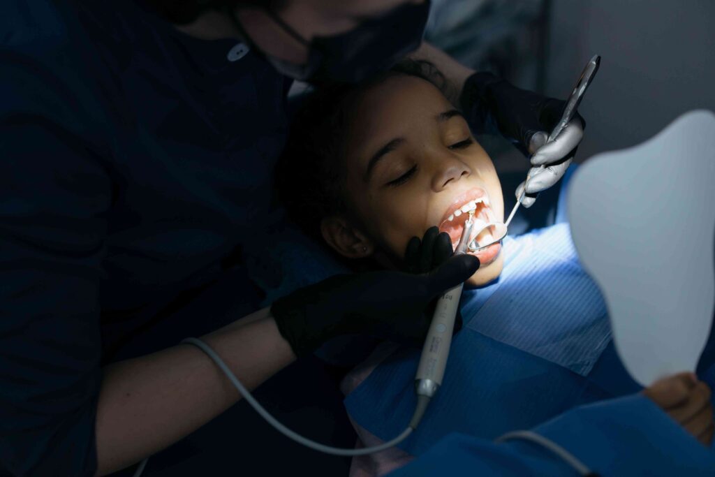
Preventive Dental Care Guide · Williamsburg Brooklyn Wondering whether dental sealants are right for your child — or for you? Here’s a clear, practical guide covering cost, safety, the procedure, insurance, and how sealants compare to fillings. Dental Sealants Cost & Safety: What Brooklyn Patients Should Know SEE AT A GLANCE SEALANTS VS FILLINGS Written by Toothology Editorial Team | Reviewed by Dr. Ayala Lieberman, DDS July 2026 If you are wondering about dental sealants — what they cost, whether they are safe, and whether they are actually necessary for your child or yourself — this guide covers everything you need to know before your next preventive visit. Sealants are not needed for everyone, but for patients with deep molar grooves or higher cavity risk, they are one of the most practical preventive tools available. A dentist will consider tooth shape, cavity risk, age, oral hygiene, existing decay, and previous sealants before recommending them. Cost depends on the number of teeth treated, insurance coverage, and visit type. Dental Sealants at a Glance What They Are Thin protective coatings applied to the chewing surfaces of back teeth — especially molars — to help reduce cavity risk in deep grooves and pits. Purpose Seal off grooves and pits where plaque and bacteria can hide — areas that are hard to reach with a toothbrush. Procedure Quick and usually painless. No drilling or numbing needed for preventive placement on healthy teeth. Who May Benefit Children and adults with deep molar grooves or higher cavity risk — especially when placed before a cavity has a chance to develop. Cost Varies by number of teeth, insurance coverage, and visit type. Often covered for children’s permanent molars under many dental plans. Longevity Can last for several years. Should be checked at routine dental visits — can be repaired or replaced if chipped or worn. How Much Do Dental Sealants Cost? The cost of dental sealants is calculated per tooth. The total depends on how many teeth are sealed, the patient’s age, insurance coverage, and whether sealants are placed during a cleaning or preventive visit. Without insurance, sealants are often a lower-cost preventive option compared with treating a cavity later. Toothology does not list a fixed sealant price because each estimate depends on the number of teeth, insurance status, and services included in the visit. GETTING YOUR COST ESTIMATE To understand your expected cost, ask Toothology to check your benefits before the appointment or request an estimate when scheduling. The office can confirm which teeth may benefit from sealants, whether they are covered, and what your out-of-pocket amount may be. What Affects the Cost of Sealants? Number of teeth being sealed in the same visit Whether the patient is a child or adult — age limits apply to many plans Insurance coverage and preventive benefits included in the plan Whether sealants are placed during a cleaning and exam or a separate visit The condition of the teeth — a sealant cannot be placed over existing decay A SEALANT CANNOT GO OVER A CAVITY If decay is found during the exam, the dentist may recommend a filling or another treatment first. This is why a cavity check is a standard part of deciding whether sealants are appropriate — and why a dental exam is always part of the visit. Are Dental Sealants Safe? For most patients, dental sealants are safe when placed by a dental professional. They are a well-established preventive treatment with a long history of use in both children and adults. Some patients ask about BPA in sealant materials. Small trace amounts may be present in some products, but dental sealants have a long and well-established history of safe use. If BPA exposure is a concern for you or your child, ask the dentist which sealant material is used. Sealants work best when placed on a clean, decay-free tooth. A dentist will check the tooth first to confirm a sealant is appropriate before proceeding. WITH A SEALANT Deep grooves and pits are sealed off — bacteria and food particles cannot collect where brushing can’t easily reach WITHOUT A SEALANT Deep grooves remain open — plaque and bacteria accumulate in the pits, increasing cavity risk on the chewing surface What Happens During the Sealant Procedure? The dental sealant procedure is quick, painless, and non-invasive. When placed on healthy teeth for prevention, numbing and drilling are not needed. Each tooth usually takes only a few minutes, and multiple teeth can be sealed in the same visit. 1 EXAM & CLEANING The dentist checks the tooth for decay or existing sealants and cleans the chewing surface before proceeding. 2 DRYING THE TOOTH The tooth is kept dry so the sealant can bond to the surface properly. 3 PREPARING THE SURFACE A gentle conditioning solution helps the sealant attach securely to the enamel. 4 APPLYING THE SEALANT The liquid sealant is painted into the grooves and pits of the chewing surface. 5 CURING A small curing light hardens the sealant in seconds. 6 BITE CHECK The dentist checks that the sealant does not affect how the teeth come together. Most patients eat and drink normally afterward. Dental Sealants vs Fillings: What Is the Difference? Sealants and fillings are not interchangeable. Sealants are preventive — placed before a cavity forms. Fillings are restorative — used to repair a tooth after decay has already developed. Factor Dental Sealant Filling Purpose Helps prevent cavities in grooves and pits on healthy teeth Repairs tooth structure after decay or damage has occurred When Used Before a cavity forms — while the tooth is still healthy After a cavity or damage is found on exam or X-ray Procedure Clean, prepare, paint on, and cure — usually no drilling or numbing Remove decay, place filling material, shape, and cure Tooth Structure Minimal to none removed from the tooth Decayed tooth structure must be removed before filling Can It Replace the Other? Cannot repair an existing cavity Cannot protect grooves like a preventive sealant THE VALUE OF PLACING SEALANTS BEFORE A CAVITY STARTS A