
Dental X-Rays in Brooklyn: How Often Do You Really Need Them?
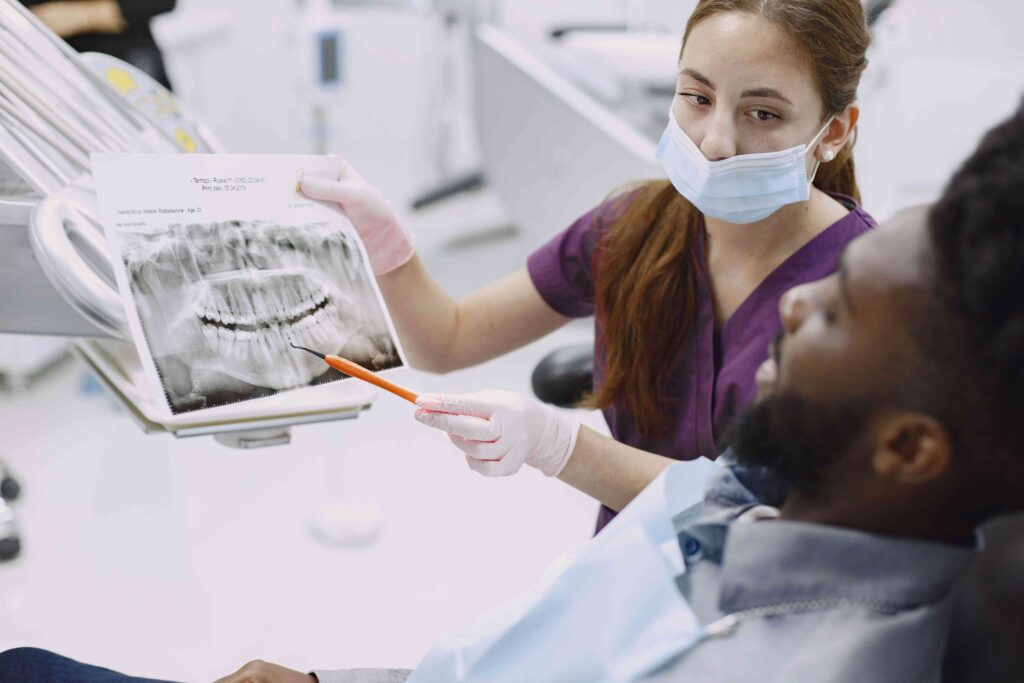
Patient Education Guide · Williamsburg Brooklyn No universal schedule applies to every patient. How often dental X-rays are recommended depends on your oral health, cavity risk, symptoms, and whether recent images are already available. Dental X-Rays in Brooklyn: How Often Do You Really Need Them? X-RAYS AT A GLANCE HOW OFTEN BY SITUATION Written by Toothology Editorial Team | Reviewed by Dr. Ayala Lieberman, DDS August 2026 Do you really need dental X-rays at every visit? Are they safe? These are reasonable questions. There is no single schedule that applies to every patient. How often dental X-rays are recommended depends on your age, oral health history, cavity risk, gum health, symptoms, and whether recent usable images are already available. X-rays help dentists see what a visual exam cannot always show — and they are used to support diagnosis, not as an automatic step at every appointment. Dental X-Rays at a Glance How Often Depends on oral health, symptoms, cavity risk, history, and whether recent usable images are already available — not a fixed schedule. Why Dentists Take Them To check for cavities between teeth, bone loss, infections, impacted teeth, and hidden issues a visual exam cannot fully see. Safety Dental X-rays use low radiation and should be recommended when they provide useful diagnostic information — following the ALARA principle. Digital X-Rays Faster, easier to store and share, and often lower in radiation than older film methods. Toothology uses digital imaging for all X-rays. BRINGING RECENT X-RAYS FROM ANOTHER OFFICE If you are visiting Toothology for the first time, bring or send any recent dental X-rays from your previous dental office. If they are current and clear, they may reduce or eliminate the need for new images at your first visit. Why Dentists Take Dental X-Rays A dental exam and a dental X-ray work together, but they do different things. During the exam, the dentist checks what can be seen directly — your gums, visible tooth surfaces, bite, soft tissue, and jaw function. X-rays show what cannot be fully seen from a visual exam alone. Cavity Detection Finding small cavities between teeth before they become larger — earlier detection usually means simpler, more conservative treatment. Bone Level Monitoring Checking for bone loss around tooth roots related to gum disease — bone changes are invisible to a visual exam. Tooth Infections Identifying infection or abscesses near the tooth root — often present without obvious visible signs. Development & Eruption Checking how children’s permanent teeth or wisdom teeth are developing and whether intervention may be needed. Treatment Planning Supporting care such as extractions, root canals, implants, or orthodontic treatment — where spatial detail matters. Hidden Problems Detecting issues beneath crowns, fillings, or existing dental work that cannot be evaluated by surface examination alone. X-RAYS SUPPORT THE EXAM — THEY DON’T REPLACE IT X-ray findings are reviewed alongside your symptoms, dental history, and exam results. An X-ray showing a finding the dentist cannot confirm clinically still leads to a careful, considered approach — not automatic treatment. How Often Do Most Patients Need Dental X-Rays? Two patients at the same practice may receive different X-ray recommendations. One may need bitewing X-rays; another may not. The decision is based on individual clinical need — not a default time-based schedule. Patient Situation What Imaging May Look Like 🆕 New Patient — No Recent X-Rays Baseline images may be needed if no current, clear records are available from a previous dental office ✅ Low-Risk Adult — Stable, No Symptoms, Recent Usable Images X-rays may be needed less often when recent images are usable and no new symptoms or concerns are present ⚠️ Higher Cavity Risk or History of Frequent Decay Bitewing X-rays may be recommended more often to monitor for new decay between appointments 📉 Gum Disease or Bone Loss History X-rays may help monitor bone levels and periodontal changes over time 🔍 Pain, Swelling, Trauma, or Suspected Infection Targeted diagnostic X-rays may be needed to find the cause — regardless of when the last ones were taken 🧒 Children and Teens Timing depends on age, tooth development, cavity risk, and exam findings — not an automatic schedule If you’re a new patient at Toothology, bring or send recent X-rays from your previous office before your visit — clear, current images may reduce or eliminate the need for new ones. Are Dental X-Rays Safe? Yes. Dental X-rays are generally safe when taken for a clear clinical reason. Modern digital dental X-rays use low radiation exposure, and dentists aim to keep that exposure as low as reasonably possible. ALARA “As Low As Reasonably Achievable” — the guiding principle for dental radiation Taking X-rays only when clinically needed Using the fewest images necessary Using digital imaging when appropriate Limiting exposure with proper technique Explaining why the X-ray is recommended Not taking X-rays simply because time has passed IF YOU ARE PREGNANT OR MAY BE PREGNANT Let the dental team know before any X-rays are taken. The dentist can review your situation, discuss whether imaging is clinically necessary, and follow appropriate safety guidance for your care. Digital X-Rays vs Traditional Film X-Rays Digital dental X-rays are used at Toothology because they offer practical benefits for both patients and dentists. Even so, the same clinical judgment applies — X-rays should still be taken only when they are clinically needed, regardless of the technology. Digital X-Rays Often lower radiation than traditional film Images appear on-screen immediately for review No chemical processing or film developing Easy to store in the patient record and share when needed Can be zoomed, enhanced, and contrast-adjusted Traditional Film X-Rays Higher radiation exposure compared with digital Require chemical processing before viewing Physical film storage, harder to transfer between offices Cannot be digitally enhanced after processing Less common in modern practices What Factors Affect X-Ray Frequency? Dentists recommend X-rays based on your current oral health, history, symptoms, and exam findings — not a default schedule. These are the factors that most commonly influence the recommendation. Age and development: Children and teens
Fluoride Treatment for Sensitive Teeth: Can It Help Enamel and Tooth Pain?

Preventive Dental Care Guide · Williamsburg Brooklyn Cold water, ice cream, or cool air causing a sharp zing? Fluoride treatment may help — but only if the right cause is identified first. Here’s what it can and can’t do, and when sensitivity means something more. Fluoride Treatment for Sensitive Teeth: Can It Help Enamel & Tooth Pain? FLUORIDE AT A GLANCE WARNING SIGNS TO WATCH Written by Toothology Editorial Team | Reviewed by Dr. Ayala Lieberman, DDS July 2026 If cold water, ice cream, or cool air causes a sharp zing in your teeth, you may be wondering whether fluoride treatment for sensitive teeth can help. The answer is: it may, depending on the cause. Fluoride can help strengthen remaining enamel and may reduce certain types of tooth sensitivity — but sensitivity can also come from cavities, cracked teeth, gum recession, exposed roots, worn enamel, infection, or failing dental work. Each cause needs a different approach. That is why the first step is a dental exam. A dentist can identify what is causing the sensitivity and recommend the right treatment — whether that is fluoride, a filling, a gum evaluation, a night guard, or another solution. Fluoride for Sensitive Teeth at a Glance What It May Help With Enamel support, early remineralisation, cavity prevention, and some dentin sensitivity linked to enamel thinning or mild gum recession. What It Cannot Fix Cavities, cracked teeth, infection, deep decay, failing restorations, or significant gum disease. These need specific dental treatment. Who May Benefit Adults and children with cold sensitivity, enamel wear, exposed roots, dry mouth, acidic diet, frequent cavities, or higher cavity risk. When to See a Dentist New, worsening, one-sided, or lingering sensitivity; pain when biting; or any sensitivity with swelling, visible damage, or fever. Why Teeth Become Sensitive Tooth sensitivity often happens when enamel becomes thin or gums recede, exposing the dentin underneath. Dentin has tiny tubules connected to the tooth nerve — so cold, heat, pressure, or touch can cause a sharp or achy response. But the cause is not always the same, even when the sensation feels similar. Possible Cause Common Pattern What May Help Enamel Erosion Cold or sweet sensitivity that may worsen over time Fluoride, remineralisation support, dietary changes, and exam Gum Recession / Exposed Roots Sensitivity near the gumline Gum evaluation, fluoride, and desensitising care Cavity or Decay Sensitivity in one tooth, often worsening with sweet or hot foods Filling or other dental treatment — fluoride cannot close a cavity Cracked Tooth Sharp pain when biting or localised sensitivity in one tooth Dental evaluation and possible restoration Teeth Whitening Temporary sensitivity during or after whitening treatment Often improves on its own; fluoride or desensitising products may help Grinding or Clenching General sensitivity, worn edges, jaw soreness Night guard evaluation; fluoride may support enamel but does not stop grinding SAME SENSATION, DIFFERENT CAUSES Sensitivity can feel similar even when the underlying cause is very different. A dental exam is the best way to find out whether fluoride is appropriate — or whether another treatment is needed first. How Fluoride Helps Protect Enamel Fluoride is a mineral that helps protect teeth against acid attacks that can weaken enamel and lead to cavities. It supports stronger enamel and can help remineralise early weakened areas before they progress into cavities. A professional fluoride treatment is significantly stronger than regular toothpaste — it is applied directly to the teeth during a dental visit and may be recommended for patients with tooth sensitivity, early enamel weakening, higher cavity risk, dry mouth, or enamel erosion concerns. FLUORIDE CAN HELP Strengthen remaining enamel Support early remineralisation of weakened areas Reduce cavity risk Protect areas affected by acid exposure Support sensitivity care when enamel or dentin exposure is part of the problem FLUORIDE CANNOT Rebuild enamel that has already been physically lost from erosion or grinding Close a cavity that has already formed Fix a cracked tooth or failing restoration Treat a dental infection or abscess Stop teeth grinding or clenching Can Fluoride Reduce Tooth Sensitivity? For some patients, yes. Professional fluoride treatment may help when sensitivity is related to enamel weakness, early demineralisation, mild wear, or exposed dentin from minor gum recession. WHEN FLUORIDE IS MORE LIKELY TO HELP Fluoride may be most helpful when sensitivity affects several teeth, appears related to enamel thinning or early wear, involves exposed roots without major gum disease, or when sensitivity is mild to moderate and stable rather than focused on one tooth or worsening. WHEN FLUORIDE IS NOT THE RIGHT SOLUTION If pain is focused on one tooth, lingers after hot or cold exposure, happens when biting, or comes with swelling — the cause may be a cavity, crack, infection, or structural issue that needs dental treatment rather than fluoride alone. When Sensitivity Means Something More Serious Mild, stable sensitivity may be manageable at home with sensitive-tooth toothpaste and good oral hygiene. But certain patterns point to a deeper problem that needs a dental exam rather than toothpaste alone. Schedule an exam if you notice any of these: Sensitivity limited to one specific tooth Pain that lingers after hot or cold exposure is removed Pain that happens without a clear trigger Pain when biting down on a specific tooth Swelling near the gum or jaw A visible hole, dark spot, crack, or chip Sensitivity that started after trauma or injury Symptoms that are getting worse over time Tooth pain accompanied by fever THESE SIGNS MAY INDICATE A CAVITY, CRACK, OR INFECTION The signs above may suggest a cavity, cracked tooth, infection, gum problem, or another issue that fluoride alone cannot fix. A dental exam is the right next step — not more toothpaste or waiting it out. Fluoride vs Toothpaste for Sensitive Teeth Sensitive-tooth toothpaste and professional fluoride treatment are not the same thing — but they are not mutually exclusive either. They can work together as part of a complete preventive care routine. Option How It Helps Best For
Dental Sealants Cost and Safety: What Brooklyn Patients Should Know
Preventive Dental Care Guide · Williamsburg Brooklyn Wondering whether dental sealants are right for your child — or for you? Here’s a clear, practical guide covering cost, safety, the procedure, insurance, and how sealants compare to fillings. Dental Sealants Cost & Safety: What Brooklyn Patients Should Know SEE AT A GLANCE SEALANTS VS FILLINGS Written by Toothology Editorial Team | Reviewed by Dr. Ayala Lieberman, DDS July 2026 If you are wondering about dental sealants — what they cost, whether they are safe, and whether they are actually necessary for your child or yourself — this guide covers everything you need to know before your next preventive visit. Sealants are not needed for everyone, but for patients with deep molar grooves or higher cavity risk, they are one of the most practical preventive tools available. A dentist will consider tooth shape, cavity risk, age, oral hygiene, existing decay, and previous sealants before recommending them. Cost depends on the number of teeth treated, insurance coverage, and visit type. Dental Sealants at a Glance What They Are Thin protective coatings applied to the chewing surfaces of back teeth — especially molars — to help reduce cavity risk in deep grooves and pits. Purpose Seal off grooves and pits where plaque and bacteria can hide — areas that are hard to reach with a toothbrush. Procedure Quick and usually painless. No drilling or numbing needed for preventive placement on healthy teeth. Who May Benefit Children and adults with deep molar grooves or higher cavity risk — especially when placed before a cavity has a chance to develop. Cost Varies by number of teeth, insurance coverage, and visit type. Often covered for children’s permanent molars under many dental plans. Longevity Can last for several years. Should be checked at routine dental visits — can be repaired or replaced if chipped or worn. How Much Do Dental Sealants Cost? The cost of dental sealants is calculated per tooth. The total depends on how many teeth are sealed, the patient’s age, insurance coverage, and whether sealants are placed during a cleaning or preventive visit. Without insurance, sealants are often a lower-cost preventive option compared with treating a cavity later. Toothology does not list a fixed sealant price because each estimate depends on the number of teeth, insurance status, and services included in the visit. GETTING YOUR COST ESTIMATE To understand your expected cost, ask Toothology to check your benefits before the appointment or request an estimate when scheduling. The office can confirm which teeth may benefit from sealants, whether they are covered, and what your out-of-pocket amount may be. What Affects the Cost of Sealants? Number of teeth being sealed in the same visit Whether the patient is a child or adult — age limits apply to many plans Insurance coverage and preventive benefits included in the plan Whether sealants are placed during a cleaning and exam or a separate visit The condition of the teeth — a sealant cannot be placed over existing decay A SEALANT CANNOT GO OVER A CAVITY If decay is found during the exam, the dentist may recommend a filling or another treatment first. This is why a cavity check is a standard part of deciding whether sealants are appropriate — and why a dental exam is always part of the visit. Are Dental Sealants Safe? For most patients, dental sealants are safe when placed by a dental professional. They are a well-established preventive treatment with a long history of use in both children and adults. Some patients ask about BPA in sealant materials. Small trace amounts may be present in some products, but dental sealants have a long and well-established history of safe use. If BPA exposure is a concern for you or your child, ask the dentist which sealant material is used. Sealants work best when placed on a clean, decay-free tooth. A dentist will check the tooth first to confirm a sealant is appropriate before proceeding. WITH A SEALANT Deep grooves and pits are sealed off — bacteria and food particles cannot collect where brushing can’t easily reach WITHOUT A SEALANT Deep grooves remain open — plaque and bacteria accumulate in the pits, increasing cavity risk on the chewing surface What Happens During the Sealant Procedure? The dental sealant procedure is quick, painless, and non-invasive. When placed on healthy teeth for prevention, numbing and drilling are not needed. Each tooth usually takes only a few minutes, and multiple teeth can be sealed in the same visit. 1 EXAM & CLEANING The dentist checks the tooth for decay or existing sealants and cleans the chewing surface before proceeding. 2 DRYING THE TOOTH The tooth is kept dry so the sealant can bond to the surface properly. 3 PREPARING THE SURFACE A gentle conditioning solution helps the sealant attach securely to the enamel. 4 APPLYING THE SEALANT The liquid sealant is painted into the grooves and pits of the chewing surface. 5 CURING A small curing light hardens the sealant in seconds. 6 BITE CHECK The dentist checks that the sealant does not affect how the teeth come together. Most patients eat and drink normally afterward. Dental Sealants vs Fillings: What Is the Difference? Sealants and fillings are not interchangeable. Sealants are preventive — placed before a cavity forms. Fillings are restorative — used to repair a tooth after decay has already developed. Factor Dental Sealant Filling Purpose Helps prevent cavities in grooves and pits on healthy teeth Repairs tooth structure after decay or damage has occurred When Used Before a cavity forms — while the tooth is still healthy After a cavity or damage is found on exam or X-ray Procedure Clean, prepare, paint on, and cure — usually no drilling or numbing Remove decay, place filling material, shape, and cure Tooth Structure Minimal to none removed from the tooth Decayed tooth structure must be removed before filling Can It Replace the Other? Cannot repair an existing cavity Cannot protect grooves like a preventive sealant THE VALUE OF PLACING SEALANTS BEFORE A CAVITY STARTS A
TMJ & Jaw Pain: Could Your Headaches Be Caused by Teeth Grinding?

TMJ & Night Guard Guide · Williamsburg Brooklyn Waking up with headaches, jaw soreness, or facial tension that eases through the day? Your teeth and jaw may be part of the pattern — and a dental evaluation can help. TMJ & Jaw Pain: Could Your Headaches Be Caused by Teeth Grinding? BOOK A TMJ EVALUATION SIGNS FROM THE JAW Written by Toothology Editorial Team | Reviewed by Dr. Ayala Lieberman, DDS July 2026 If you wake up with headaches, jaw soreness, or facial tension that eases as the morning goes on, your teeth and jaw may be part of the problem. A TMJ headache can feel like pressure near the temples, jaw, ears, or face — and may be worse after a night of clenching or grinding without you even realising it is happening. Many people with recurring morning headaches never consider a dental connection. But when headaches happen alongside jaw pain, tooth wear, stiffness, or clenching, a dental evaluation can help determine whether your jaw, bite, or teeth grinding is contributing to the pattern. TMJ HEADACHES ARE ONE POSSIBLE CAUSE — NOT THE ONLY ONE TMJ headaches and bruxism are one possible cause of recurring headaches. Tension headaches, migraines, and medical conditions can cause similar symptoms. However, when jaw symptoms are part of the pattern, dentistry is worth including in the conversation. What Is TMJ and How Does It Cause Headaches? THE TEMPOROMANDIBULAR JOINT (TMJ) The TMJ is the hinge joint that connects the lower jaw to the skull, just in front of each ear. You use it every time you chew, speak, yawn, or swallow. When this joint or the surrounding jaw muscles become irritated, strained, or overworked, it can lead to temporomandibular disorders (TMD) — what most people call “TMJ problems.” Pain from the jaw area can spread toward the temples, ears, face, neck, or head. TMJ-related headaches can occur when jaw muscles are under repeated stress from clenching or grinding — especially during sleep. The jaw muscles are among the most powerful in the body, and when they work while they are supposed to be resting, the resulting muscle fatigue and tension can radiate outward toward the head, temples, and ears. When headaches occur with jaw soreness, chewing discomfort, facial tension, or pain near the ears, TMJ and teeth grinding may be worth evaluating. Can Teeth Grinding Really Cause Jaw Pain and Headaches? Bruxism — the clinical term for teeth grinding and jaw clenching — can contribute to jaw pain, facial tension, and headaches. When you clench or grind during sleep, the jaw muscles work far harder than they should while the rest of the body is resting. Over time, this leaves the chewing muscles tired, sore, and tight. Many people do not know they grind their teeth because sleep bruxism happens unconsciously. In some cases, a dentist may notice signs — such as tooth wear, jaw tenderness, or bite-related stress — before the patient has any idea grinding is occurring. Signs Bruxism May Be Affecting You Dull headaches near the temples, especially in the morning Jaw soreness, stiffness, or tight feeling when waking Facial muscle tension that eases during the day Tooth sensitivity, especially after waking Pain or pressure near the ears or jaw joint Teeth that look flat, worn, shorter, or chipped Difficulty opening the jaw wide in the morning A sleep partner who has heard grinding sounds at night TMJ Headache vs Tension Headache vs Migraine TMJ headaches, tension headaches, and migraines can overlap, which is why self-diagnosis is not always reliable. A clear pattern and a proper evaluation help identify what is driving the symptoms. Headache Type Common Pattern Clues to Look For Next Step TMJ-Related Pain or pressure near the temples, jaw, ears, face, or neck — often worse in the morning Jaw pain or soreness, clenching or grinding, chewing discomfort, clicking, morning stiffness, or visible tooth wear Dental / TMJ evaluation Tension Headache Band-like pressure around the head or forehead Neck and shoulder tension, stress, general muscle tightness — not specifically jaw-related Medical or dental evaluation depending on symptoms Migraine Moderate to severe pain, often one-sided or throbbing Light or sound sensitivity, nausea, visual disturbances, worsens with activity Medical evaluation WHEN TO SEEK URGENT MEDICAL CARE — NOT A DENTAL VISIT A sudden, severe headache unlike any previous headache Headache with fever or stiff neck Headache after significant head trauma Confusion, weakness, vision changes, or fainting Headache with chest pain or shortness of breath THESE SYMPTOMS ARE NOT TYPICAL TMJ SIGNS The warning signs above suggest medical causes that need urgent evaluation. They should not be approached as a dental issue. If you have any of these symptoms, seek medical care promptly. Signs Your Headache Is Coming From Your Jaw These signs do not confirm a TMJ diagnosis on their own — but if several apply together, a dental evaluation is a reasonable next step to see whether the jaw, bite, or teeth grinding is contributing. Morning headaches that ease as the day progresses Jaw soreness, stiffness, or pain when waking up Pain near the temples, ears, face, or jaw Clicking, popping, or grinding sounds in the jaw Pain or discomfort when chewing Awareness of clenching or grinding during the day A partner has heard grinding sounds during sleep Teeth that look flatter, shorter, chipped, or worn Tooth sensitivity without a clear cavity or gum issue Facial tension, neck stiffness, or tired jaw muscles How Dentists Diagnose TMJ-Related Headaches A dental evaluation for suspected TMJ headaches or bruxism is usually simple and non-invasive. The goal is to find out whether the teeth, bite, jaw joint, or jaw muscles may be contributing to symptoms. Symptom history: Headache timing, morning jaw pain, soreness, sleep habits, stress levels, and any grinding sounds reported by a partner Teeth: Signs of grinding — flat surfaces, chipped edges, cracks, worn enamel, or unusual sensitivity Bite: How the teeth come together and whether bite pressure may be straining the jaw joint
Oral Cancer Screening: What Dentists Check During a Routine Exam
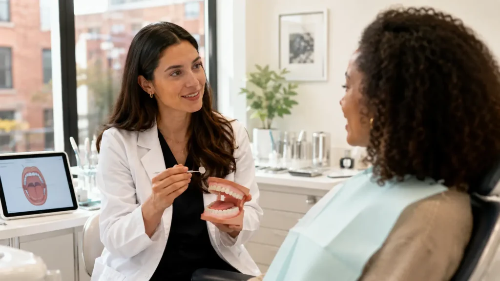
Preventive Dentistry Guide · Williamsburg Brooklyn Most people know their dentist checks for cavities. What many don’t realise is that a thorough exam also includes a soft tissue check — a visual and hands-on evaluation of the gums, tongue, cheeks, lips, and throat area. Oral Cancer Screening: What Dentists Check During a Routine Exam WHAT THE SCREENING INVOLVES SIGNS WORTH CHECKING Written by Toothology Editorial Team | Reviewed by Dr. Ayala Lieberman, DDS July 2026 Most people come to a routine dental exam expecting their dentist to check for cavities and assess gum health. What many do not realise is that a thorough dental exam also includes a soft tissue check — a visual and hands-on evaluation of the gums, cheeks, tongue, lips, floor and roof of the mouth, and the visible throat area. A red mark on the gum, a small sore that won’t heal, or a white patch noticed while brushing can all feel alarming. Most of the time, these changes have straightforward explanations — a minor irritation, a bite injury, a gum infection, or a temporary reaction. What matters clinically is not the appearance of a change in isolation, but whether it persists, worsens, or lacks an obvious explanation after a reasonable period. What Is an Oral Cancer Screening? An oral cancer screening is a quick visual and hands-on check of the mouth, gums, tongue, throat area, and nearby soft tissues. It helps identify unusual changes that may need monitoring or further evaluation. In most dental offices, this screening is part of a routine dental exam — it is painless, takes only a few minutes, and usually requires no special preparation. Part of Your Routine Exam An oral cancer screening is not a separate procedure. It is included as part of a comprehensive dental exam at Toothology in Williamsburg. Quick and Painless The check takes only a few minutes. No injections, no special equipment, and no preparation is required on your part. Screening ≠ Diagnosis A screening does not diagnose cancer. It helps your dentist spot changes that may need closer review. Only a biopsy examined by a pathologist can confirm a diagnosis. Covers Areas You Can’t See Yourself Areas like the underside of the tongue, floor of the mouth, and back of the throat are difficult to examine clearly at home — the exam provides proper lighting and positioning. Why Dentists Check the Gums, Tongue, Cheeks, and Throat A dental exam is not only about teeth. Dentists also check the soft tissues of the mouth because they can spot changes that patients may not notice on their own. During a routine exam, the dentist uses proper lighting, positioning, and touch to look for unusual tissue changes in each of these areas. Area Examined What the Dentist Looks and Feels For 👄 Lips & Lip Corners Sores, ulcers, rough areas, discolouration, swelling, or asymmetry 🫦 Cheeks & Inner Lining Red or white patches, lumps, ulcers, or thickened tissue along the inner cheek surface 🦷 Gums Persistent sores, unusual redness, white patches, swelling, or unexplained gum changes 👅 Tongue Red or white patches, lumps, ulcers, or restricted movement — top, sides, and underside are all checked ⬇ Floor of the Mouth Swelling, lumps, ulcers, or unusual tissue texture under the tongue — an area that is hard to check at home ⬆ Roof of the Mouth Persistent sores, red or white patches, or abnormal tissue changes on the hard or soft palate 🗣 Back of the Throat Visible changes or persistent unexplained irritation in the visible throat area 🫀 Jaw & Neck Enlarged lymph nodes or unexplained lumps felt during palpation of the jaw and neck area A screening does not diagnose cancer — it helps your dentist identify changes that may need monitoring or further evaluation. Only a biopsy examined by a pathologist can confirm a diagnosis. Oral Cancer Signs on the Gums That Should Be Checked The gums can show changes from irritation, infection, brushing habits, dentures, or other common causes. Not every red spot or sore is alarming — but changes that persist for about two weeks, worsen, grow, bleed without explanation, or cause numbness or pain should be checked by a dentist. A red or white patch on the gums, tongue, or inside the mouth lasting about two weeks A sore, ulcer, or rough area that does not heal or keeps returning A persistent red mark on the gum, cheek, or lip with no clear cause A lump, bump, or thickened area in the mouth, jaw, or neck Unexplained bleeding from gum or mouth tissue Numbness, tenderness, or pain without an obvious cause Difficulty chewing, swallowing, or moving the tongue or jaw Persistent sore throat or hoarseness lasting more than two weeks A loose tooth without a clear dental explanation Is a Red Mark on the Gum Always Serious? A red mark on the gum is usually caused by something benign — gum inflammation, minor irritation, infection, a canker sore, or a reaction to certain foods or dental products. Most mouth changes are not cancer. A red spot, gum sore, or patch is more often related to irritation, an accidental bite, brushing trauma, or gum disease. The Persistence Rule Usually less concerning: The red mark improves within one to two weeks — consistent with irritation, a minor bite, or temporary inflammation. Worth monitoring: The mark is present for two weeks but not worsening — worth mentioning at your next routine exam for documentation. Should be checked: The red mark improves within one to two weeks — consistent with irritation, a minor bite, or temporary inflammation. What Happens During a Dentist’s Oral Cancer Screening? During an oral cancer screening, the dentist or hygienist checks the soft tissues of the mouth and nearby areas for unusual changes. The process is calm, thorough, and completed as part of the regular exam. Face and neck: Checking for swelling, asymmetry, or enlarged lymph nodes Lips: Looking for sores, scaling, discolouration, or uneven changes Cheeks: Checking for red or white patches, ulcers, or
How Do Cavities Form? A Dentist’s Guide to Stopping Tooth Decay Early
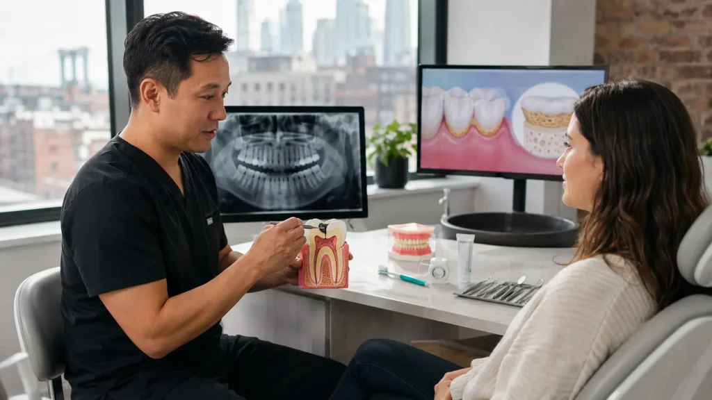
Preventive Dentistry Guide · Williamsburg Brooklyn Most cavities don’t start with pain. Understanding the predictable process behind tooth decay — plaque, bacteria, sugar, acid, and time — is the key to stopping it early. How Do Cavities Form? A Dentist’s Guide to Stopping Decay Early SEE HOW IT FORMS DAILY PREVENTION HABITS Written by Toothology Editorial Team | Reviewed by Dr. Ayala Lieberman, DDS June 2026 Most cavities do not start with pain. A dentist points to a small dark spot on an X-ray and a patient says they had no idea it was there. A teen comes in for a routine checkup and there’s a soft groove on a back molar that wasn’t there six months ago. A parent who brushes their child’s teeth carefully every night still hears that a cavity has formed. The answer to how cavities form starts with plaque, bacteria, sugar, acid, and time — and understanding that process is the foundation of stopping decay early. A cavity is not something that appears overnight. It is the end result of repeated acid attacks on tooth enamel over time, and that process gives you multiple opportunities to interrupt it before it becomes a hole that needs a filling. The earlier tooth decay is caught, the more conservative the solution can usually be. How Cavities Actually Form Tooth decay follows a specific, predictable sequence. Understanding each step makes it much clearer why certain habits accelerate decay and why certain preventive measures work. 1 PLAQUE FORMS Bacteria, food particles, and saliva create a sticky film on the teeth — this happens to everyone, every day. 2 BACTERIA FEED ON SUGAR Bacteria in plaque consume sugars and fermentable carbohydrates from the foods and drinks you consume. 3 ACID IS PRODUCED The bacteria release acids as a byproduct, and that acid attacks the enamel surface within minutes of eating or drinking. 4 ENAMEL LOSES MINERALS Repeated acid exposure weakens enamel and may create white or chalky spots — the first visible sign of demineralisation. 5 ENAMEL BECOMES POROUS Without enough saliva, fluoride, and recovery time, the weakened spot can progress into a small hole — a true cavity. 6 DECAY SPREADS INWARD Untreated decay can move into dentin and toward the nerve, causing pain or infection if it continues unaddressed. IT’S NOT SUGAR ALONE — IT’S FREQUENCY The key issue is not sugar alone — it is how often teeth are exposed to acid. Sipping a sugary drink throughout the day causes repeated acid attacks, while having the same amount of sugar at one time gives teeth more recovery time. That’s why snacking habits and drink frequency matter so much in cavity prevention. Why Plaque and Acid Damage Tooth Enamel Plaque forms on everyone’s teeth every day. It’s a sticky film of bacteria, and the problem starts when those bacteria frequently feed on sugars and refined carbohydrates. After you eat or drink anything other than water, plaque bacteria produce acid that attacks enamel within minutes. Saliva helps neutralise acid and return minerals to the tooth surface, but it needs time. When teeth are exposed to sugar or acid too often — or when saliva flow is low — enamel damage can outpace repair. Risk Factors for Enamel Erosion and Decay Incomplete Brushing & Flossing Plaque that is not removed can harden into tartar, which requires professional cleaning to fully remove. Frequent Sugar or Acid Exposure Juice, sports drinks, soda, crackers, white bread, and dried fruit can feed decay-causing bacteria, especially consumed slowly over time. Dry Mouth Reduced saliva flow from medications, health conditions, or mouth breathing increases cavity risk significantly. Deep Grooves in Molars Some back teeth have grooves that trap bacteria and food, making them harder to clean thoroughly. Orthodontic Appliances Braces, wires, and aligners create more places for plaque to collect and hide from a toothbrush. Missed Dental Cleanings Professional cleanings remove tartar and help reduce the bacterial buildup that contributes to decay over time. Early Signs of a Cavity Many early cavities cause no pain at all. Discomfort often appears only after decay has moved deeper into the tooth, which is why routine exams and X-rays are important. That said, these signs are worth mentioning at your next dental visit. A white, chalky, or matte spot on the tooth surface A dark brown or black spot in a groove or pit Sensitivity to sweets or cold drinks A visible pit, rough edge, or small depression Food getting stuck in the same spot repeatedly Floss catching or tearing in one specific area A bad taste from one part of the mouth A mild, dull ache in a specific tooth THESE SIGNS DON’T CONFIRM A CAVITY — BUT THEY CONFIRM AN EXAM These signs do not always mean you have a cavity. Stains, sensitivity, and rough edges can have other causes. But an exam and X-rays can confirm what’s happening and help keep treatment more conservative if decay is found. Can a Cavity Heal on Its Own? It depends on the stage of decay. If you’re asking “can you heal a cavity?” or “will cavities heal?”, the answer depends on whether decay is still in the early enamel mineral-loss stage or whether a true hole has already formed. EARLY STAGE — BEFORE A HOLE FORMS Enamel mineral loss can sometimes be slowed, stopped, or partially reversed Better brushing and fluoride toothpaste can help strengthen enamel Reduced sugar frequency gives teeth more recovery time Professional fluoride treatment can accelerate remineralisation TRUE CAVITY — HOLE HAS FORMED Cannot heal or close on its own — teeth do not regrow lost structure Dental treatment is needed, usually a filling Waiting allows decay to spread deeper into the tooth Home remedies cannot reverse an actual structural hole A NOTE ON OIL PULLING AND HOME REMEDIES Searches like “oil pulling teeth cavity” often overstate what oil pulling can do. Oil pulling is sometimes promoted online for cavities, but it should not replace brushing with fluoride toothpaste, flossing, or professional dental care. It
All-on-4 Dental Implants vs. Dentures: Which Is the Better Investment?

Full-Arch Restoration Guide · Williamsburg Brooklyn An honest comparison — stability, bone health, cost of ownership over 10 years, and who is the right candidate for each option. No overselling either direction. All-on-4 Dental Implants vs. Dentures: Which Is the Better Investment? SEE THE FULL COMPARISON WHO IS THE RIGHT CANDIDATE? Written by Toothology Editorial Team | Reviewed by Dr. Ayala Lieberman, DDS April 2026 If you are living with dentures — or facing the prospect of significant tooth loss — you have almost certainly heard the term “All-on-4 dental implants” come up in conversation, in advertising, or in a consultation room. The honest answer is that dentures vs. implants is not a simple better/worse question. It depends on your bone health, medical history, financial situation, and what you are actually hoping to change about your current experience. Neither option should be approached as a one-size-fits-all decision. Dentures can be the right solution for many patients — particularly where surgical contraindications exist or where cost is the overriding constraint. All-on-4 is a surgical procedure with a recovery process and specific candidacy requirements. What follows is an honest account of both. The Difference Between Removable Dentures and All-on-4 Implants The most fundamental distinction between the two options is fixed versus removable. Removable dentures sit on top of the gum tissue and are held in place by suction, clasps on remaining teeth, or dental adhesive. They come out at night, require soaking and cleaning in a separate container, and rely on the continued fit of the gum tissue beneath them — which changes as the underlying bone resorbs over time. All-on-4 is a full-arch implant concept in which the entire replacement arch of teeth is supported by four implants placed strategically in the jawbone. The prosthetic — often a zirconia bridge or acrylic hybrid on a titanium framework — is fixed to the implants and does not come out for daily cleaning. It does not require adhesive, does not rely on gum suction, and because the implants are embedded in bone, they stimulate it in a way that removable dentures do not. NOTE ON “PERMANENT DENTURES” This term is used inconsistently — it sometimes refers to dentures that are implant-supported and non-removable, and sometimes to high-quality conventional dentures intended for long-term use. A fixed implant-supported arch is more accurately described as a full-arch implant bridge rather than a denture in the conventional sense. Full Side-by-Side Comparison Category Removable Dentures All-on-4 Implants Daily Stability Relies on suction, adhesive, or clasps; can shift during speaking and eating Fixed to implants; does not move, slip, or require adhesives during the day Bite Force Significantly reduced compared to natural teeth; hard foods often avoided Substantially stronger than removable dentures; allows a wider range of foods (case-dependent) Jawbone Preservation Does not stimulate bone; continued resorption changes face shape and denture fit over time Implants stimulate bone similarly to natural tooth roots, helping to slow ongoing resorption Daily Care Removed nightly, soaked, brushed; adhesive reapplied each morning Cleaned in place with soft brush, floss tools, water flosser; removed only by clinician Adjustments & Repairs Periodic relines needed as bone changes; repairs for cracks or fractures; replacement over time Periodic check-ups; prosthetic may need adjustment or replacement over many years; implants typically remain Comfort Over Time Fit can worsen as bone shrinks; sore spots and gum irritation are common complaints No gum pressure from a removable plate; discomfort during healing period is temporary Speech Initial adjustment period; clicking or slipping can affect speech, particularly without adhesive Fixed prosthetic closely mimics natural tooth position; most patients adapt quickly Long-Term Appearance Facial appearance may change over time as underlying bone is resorbed Implants help preserve bone contour, supporting a more stable facial profile over time Why All-on-4 Is Often Called “Teeth in a Day” The phrase “Teeth in a Day” — sometimes described as same-day implants or immediate loading — refers to the possibility of placing implants and attaching a temporary fixed prosthetic within a single surgical appointment. The concept involves strategically angling implants — often tilting the posterior implants to avoid the sinus cavity and areas of reduced bone density — to maximise contact with available bone. WHAT “SAME DAY” ACTUALLY MEANS “Same day” refers to the temporary bridge, not the final bridge. The final prosthetic comes after osseointegration is confirmed — typically several months later. And not every patient qualifies for immediate loading — this is determined at consultation with 3D imaging. What the Full Treatment Process Typically Involves 1 CONSULTATION & 3D IMAGING A thorough examination including CBCT (3D cone beam CT) imaging to assess bone volume, bone density, nerve positions, sinus anatomy, and overall jaw suitability for implant placement. 2 TREATMENT PLANNING Reviewing options for number of implants, prosthetic materials (zirconia bridge, acrylic hybrid), timeline, and any prerequisite treatment needed — including extractions or bone grafting. 3 EXTRACTIONS (IF NEEDED) Remaining failing teeth are extracted — often on the same day as implant placement in qualifying cases, reducing the total number of surgical appointments. 4 IMPLANT PLACEMENT Implants are surgically placed into the jawbone under local anaesthesia, with sedation options available. Implants are tested for primary stability before any immediate loading decision is made. 5 TEMPORARY FIXED BRIDGE (SAME DAY — IF QUALIFIED) In cases where sufficient primary stability is achieved, a temporary prosthetic is attached to the implants on the same day — allowing you to leave with fixed, functional teeth. 6 HEALING & OSSEOINTEGRATION Over several months, the implants fuse with the surrounding bone. Regular monitoring appointments assess integration progress and ensure healing is on track. 7 FINAL PROSTHETIC Once osseointegration is confirmed, the temporary bridge is replaced with the final restoration — often a full-arch zirconia bridge or similar durable prosthetic designed for long-term wear. Long-Term Benefits: Bone Health and Dietary Freedom Jawbone Preservation: Why It Affects Your Face, Not Just Your Teeth When natural teeth are lost, the bone that previously surrounded their roots no longer receives the stimulation it needs to maintain its density and volume. Without that stimulation,
Why Are My Teeth Suddenly Sensitive to Cold Water? Causes and Treatment

Tooth Sensitivity Guide · Williamsburg Brooklyn That sharp zing when you sip cold water isn’t just annoying — it’s a signal. Cold sensitivity is a symptom, not a diagnosis. Here’s what’s actually behind it and what fixes it. Why Are My Teeth Suddenly Sensitive to Cold Water? Causes & Treatment FIND THE CAUSE SEE THE TREATMENTS Written by Toothology Editorial Team | Reviewed by Dr. Ayala Lieberman, DDS June 2026 You take a sip of iced coffee and feel a sharp zing in your tooth. Cold air hits your teeth on a winter morning and you wince. A glass of cold water suddenly feels different than it did a few months ago. Cold sensitivity is one of the most common dental complaints — but also one of the most misunderstood. Some people assume it’s normal and reach for a desensitising toothpaste. Sometimes that helps. Other times, it only masks a bigger issue like a cavity, crack, gum recession, or failing filling. The key thing to understand is that cold sensitivity is a symptom, not a diagnosis. Mild sensitivity across several teeth is different from sharp, lingering pain in one tooth — and those differences matter when deciding what to do next. What Triggers Sharp, Sudden Temperature Sensitivity? When cold water causes a sharp zing, the problem is usually not the enamel itself. Enamel is the hard outer layer of the tooth and does not contain nerves. Beneath it is dentin, which contains tiny channels called dentinal tubules that connect toward the inner nerve of the tooth. When dentin becomes exposed — from enamel wear, gum recession, a crack, or a cavity — cold water or air can trigger movement inside those tubules. That movement stimulates the nerve and creates the quick, sharp pain people describe as tooth sensitivity. UNDERSTANDING TOOTH LAYERS — WHY COLD REACHES THE NERVE ENAMEL — OUTER SHELL The hardest substance in the body. Contains no nerves. When intact, it fully insulates the dentin and pulp. When worn or eroded, dentin sits closer to the surface. DENTIN — THE SENSITIVE LAYER Contains dentinal tubules — microscopic channels running toward the nerve. Exposed dentin is the direct cause of cold sensitivity. Root surfaces have no enamel — only thinner cementum — making them especially reactive. PULP & NERVE — INNER CORE Soft tissue containing the tooth’s nerve supply. Pain that lingers several seconds after the cold trigger is gone may suggest the nerve is significantly involved. WHY COLD WATER HURTS MORE THAN ROOM-TEMPERATURE WATER Cold water triggers a stronger response because it causes a faster fluid shift inside exposed dentinal tubules. That sudden movement stimulates the nerve and produces the sharp, immediate pain people notice. The same response can happen with cold air — particularly when root surfaces or worn enamel are exposed. Acidic foods and drinks can make this temporarily worse by softening the surface and opening tubules further. WHY “SUDDEN” SENSITIVITY MATTERS When sensitivity feels sudden or is noticeably worsening, it often means something has changed. Gum recession may have exposed more root surface. Enamel may have worn further. A crack may have formed. Or a filling may no longer be sealing the tooth properly. Sensitivity that appears recently or escalates quickly deserves more attention than sensitivity that has stayed mild and stable over a long period. How Gum Recession Uncovers Tooth Nerve Paths Enamel only covers the crown of the tooth — the visible portion above the gumline. Below the gumline, the root surface is covered by cementum: a thinner, far less protective material. When gum tissue recedes — pulling away from the tooth and exposing part of the root — that exposed surface produces a much more pronounced sensitivity response than comparable dentin exposure on the crown. Gum recession is one of the most common causes of cold sensitivity. It often develops slowly — which is why the pain can feel sudden once enough root surface finally becomes exposed. Aggressive Brushing Brushing too hard or using a medium or hard-bristle toothbrush can gradually wear the gumline and expose sensitive root surfaces over months to years. Abrasive Toothpaste Some whitening or “heavy-cleaning” toothpastes can increase gumline wear — particularly when combined with forceful brushing technique. Periodontal Disease Gum disease causes the gums and supporting bone to pull away from the tooth, leading to more significant recession — often across multiple teeth simultaneously. Naturally Thin Gum Tissue Some people naturally have thinner gum tissue, which makes recession more likely over time regardless of hygiene habits. PRODUCT CHANGES AREN’T ENOUGH Switching to a softer toothbrush and less abrasive toothpaste can help prevent further damage — but it won’t restore gum tissue that has already receded, address gum disease if that’s the cause, or tell you how severe the recession is or whether it’s still progressing. A dental evaluation is more useful than relying on a product change alone. In-Office Desensitising vs. Store-Bought Sensitivity Toothpaste Sensitive teeth treatments range from over-the-counter toothpaste to in-office desensitising procedures. The right option depends on the cause and severity of the sensitivity. When Sensitivity Toothpaste Is a Reasonable Starting Point Sensitivity toothpastes with ingredients like potassium nitrate or stannous fluoride can help reduce mild, generalised sensitivity by calming the nerve response or blocking exposed dentinal tubules. They can be a reasonable first step for minor enamel wear or temporary post-whitening sensitivity. THE LIMITATION OF TOOTHPASTE ALONE Toothpaste only helps manage the symptom. It does not treat the underlying cause if the problem is gum recession, a crack, a failing filling, or early decay. If sensitivity is worsening, localised, or persistent — relying on toothpaste alone can delay the right diagnosis and allow the underlying issue to worsen. How Treatment Options Compare Treatment Option What It Does Best Suited For At-Home Sensitivity Toothpaste Potassium nitrate / stannous fluoride Calms nerve endings in dentin tubules over repeated use; gradual improvement over several weeks of consistent use Mild diffuse dentin hypersensitivity; ongoing maintenance for sensitivity-prone patients; first step for minor enamel wear In-Office Fluoride Treatment or Varnish Concentrated mineral application
Wisdom Teeth Removal in Williamsburg: How 3D Imaging Makes Surgery Safer
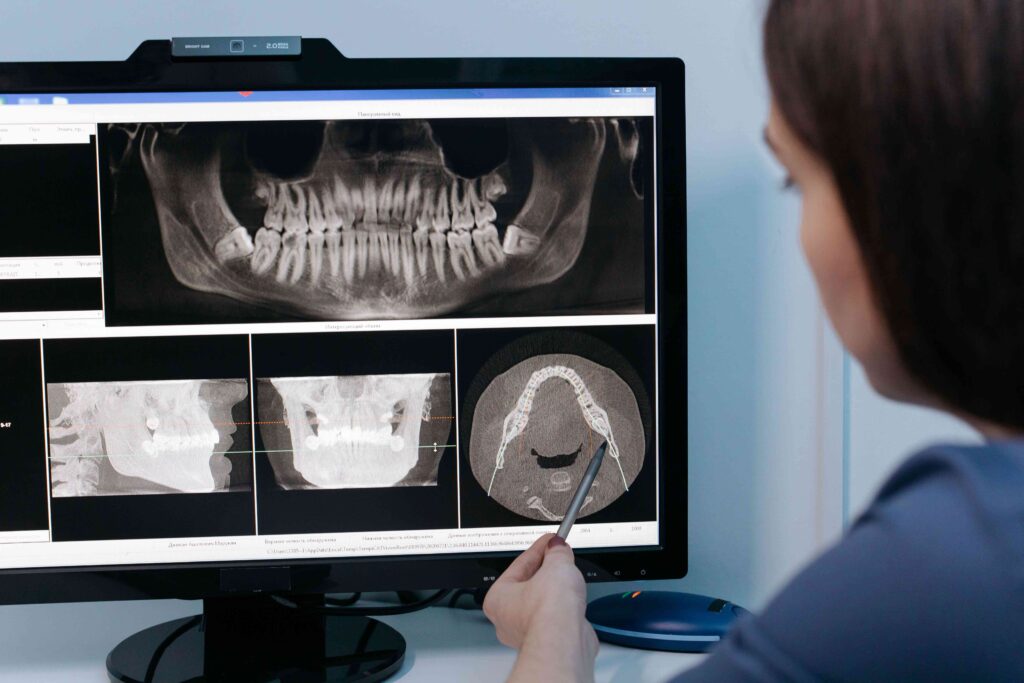
Wisdom Teeth Removal Guide · Williamsburg · 3D Imaging CBCT 3D imaging shows the exact path of the nerve, root shape, and jaw anatomy that a flat 2D X-ray can’t. Here’s why it changes the surgical plan — and what recovery actually looks like. Wisdom Teeth Removal in Williamsburg: How 3D Imaging Makes Surgery Safer HOW 3D IMAGING HELPS THE 48-HOUR RECOVERY PLAN Written by Toothology Editorial Team | Reviewed by Dr. Ayala Lieberman, DDS June 2026 For most people, wisdom teeth removal is something they know is coming long before it becomes urgent. A dentist mentions it at a routine checkup. An X-ray shows third molars heading in the wrong direction. Or the jaw starts aching in a way that feels different from anything before. Then comes the question most people delay longer than they should: when is the right time, and how do I make sure this goes as smoothly as possible? Wisdom teeth removal is one of the most commonly performed dental procedures, and for the vast majority of patients it goes without significant complication. But the cases that generate the most anxiety, and the most serious post-operative concerns, often involve anatomy that isn’t fully characterised on initial 2D imaging — especially when roots appear close to important nerves. Modern CBCT 3D imaging has changed that picture significantly. Why 3D Nerve Mapping Is Essential for Safe Third Molar Extraction The inferior alveolar nerve — which supplies sensation to the lower lip, chin, and lower teeth — runs in a canal in the lower jaw near the roots of lower wisdom teeth. In simple, fully erupted cases, a panoramic X-ray may be enough. But with deep impactions or roots that appear close to the nerve on a 2D image, there can be real uncertainty. A CBCT (cone beam CT) 3D scan gives a full three-dimensional view of the jaw so the surgeon can see the exact relationship between the roots and the nerve canal — information that can directly affect the surgical plan. Whether to Section the Tooth Removing the tooth in smaller pieces can reduce force near nerve-adjacent roots. CBCT helps guide that decision more precisely than a flat image can. Which Approach to Use 3D mapping can influence the entry angle, how much bone needs to be removed, and the safest direction of controlled pressure during extraction. Whether to Refer CBCT may reveal complexity that is better handled by a specialist — far better to know before surgery than to discover it mid-procedure. Sinus Proximity (Upper Teeth) For upper wisdom teeth, 3D imaging maps the floor of the maxillary sinus precisely — helping avoid sinus communication during extraction. IMPORTANT CLARIFICATION: IMAGING REDUCES RISK — IT DOES NOT ELIMINATE IT A CBCT scan is a planning tool, not a guarantee. If a wisdom tooth sits close to the inferior alveolar nerve, there is still some risk of temporary tingling or numbness in the lower lip or chin. In most cases, these sensory changes improve as the nerve heals, and permanent injury is rare. What CBCT does is give the clearest possible view of your anatomy so the surgeon can plan the most precise, nerve-aware approach. What CBCT Shows That a Panoramic X-Ray May Not Imaging Capability Panoramic X-Ray CBCT 3D Scan View Dimension 2D flat image — one perspective only Full 3D volumetric view from any angle Nerve Path Visualisation General proximity only — nerve appears as a line on the image Precise 3D path of the inferior alveolar nerve relative to each root tip Root Shape & Anatomy Overlapping roots can be ambiguous or misleading on a flat image Each root tip and its exact position shown clearly in three dimensions Depth & Angulation Limited — bucco-lingual (front-to-back) depth cannot be assessed Full depth and exact angulation of each root visible in all planes Adjacent Structure Mapping Maxillary sinus proximity is only approximate Sinus floor, adjacent teeth, and bone density all visible and measurable Surgical Planning Impact Sufficient for straightforward, fully erupted teeth Enables modified surgical approaches for high-risk nerve proximity cases Radiation Level Lower dose; commonly used as the initial screening image Higher than panoramic but typically lower than medical CT; used when 3D detail is clinically warranted A CBCT scan is a planning tool, not a guarantee. It gives the clearest possible view of root-to-nerve relationships so the surgeon can plan the most precise, nerve-aware approach — but some risk remains even with optimal imaging. Symptoms Indicating Your Wisdom Teeth Are Damaging Adjacent Molars Wisdom teeth don’t have to be fully impacted to cause problems. Even partially erupted or misaligned third molars can generate a constellation of symptoms that, if left unaddressed, lead to damage extending well beyond the wisdom tooth itself. Jaw pain or persistent deep aching — often worse at night or when chewing Swelling or tenderness in the back gum — pericoronitis that keeps coming back Bad taste or bad breath in one spot — food and bacteria trapped behind a partially erupted tooth Headaches or jaw tension — impaction pressure can contribute to jaw and temple headaches Repeated gum infections — antibiotics may calm a flare-up, but recurrence usually means extraction Damage to the second molar — impacted wisdom teeth can cause decay or root resorption on the adjacent molar Bone loss near the second molar — X-rays may show bone breakdown or deep pocketing between the two teeth Difficulty or discomfort opening the mouth — especially with swelling or infection present WHEN THE DECISION CANNOT WAIT Watching an impacted wisdom tooth can be reasonable when it is truly symptom-free — but that window often closes once the second molar starts showing bone loss or root resorption. That can become a bigger problem than the extraction itself. Go to the ER or Urgent Care Immediately If You Have: Swelling spreading to the throat, neck, or floor of the mouth Difficulty breathing or swallowing High fever with significant facial swelling
Should You Get a Second Opinion on Your Dental Treatment Plan?

Patient Guidance · Treatment Planning When a treatment plan feels bigger than expected, a second opinion is not distrust — it is the most rational way to approach a decision that is significant in cost, impact, or permanence. Should You Get a Second Opinion on Your Dental Treatment Plan? Written by Toothology Editorial Team | Reviewed by Dr. Ayala Lieberman, DDS May 2026 You walk out of a dental appointment holding a treatment plan that lists procedures you were not expecting — several crowns, a root canal, possible extractions, or a full-mouth reconstruction that comes with a price tag that makes you hesitate at the door. You trust your dentist well enough. But something about the conversation felt rushed, or the scale of the recommended work surprised you, or you simply want to understand your options before committing to something this significant. Seeking a dental second opinion in that situation is not an act of distrust. It is a reasonable, patient-centred step that any responsible dental professional should welcome. Dental Emergency? If you are dealing with a dental emergency — active infection, significant swelling, severe pain — please seek evaluation promptly rather than waiting for a second opinion to be arranged. Why High-Cost Dental Work Often Warrants a Second Look There is nothing inherently suspicious about receiving a large treatment plan from a dentist. Dental disease is progressive, and it is genuinely common for patients who have deferred care for a few years to discover multiple problems at once. Situations Where a Second Opinion Is Particularly Worth Pursuing You have been quoted for five or more crowns, implants, or major restorations at once The consultation felt rushed and you left without a clear understanding of what is wrong Created with Sketch Beta. You have been recommended for full-mouth rehabilitation, All-on-X, or a significant bite reconstruction You have been told to extract a tooth you were not expecting to lose — without a clear explanation The treatment plan includes procedures you have not been given alternatives for Recommendations have changed significantly between dentists without clear explanation You feel pressure to commit quickly or offered heavy discounts for same-day decisions A large root canal or surgical procedure is recommended and you want to understand the basis Urgency Changes the Calculation Not every dental situation can wait. Swelling, spreading infection, significant pain, or any sign of a dental abscess are situations where prompt evaluation and treatment are clinically appropriate. If your dentist has expressed any urgency around a treatment recommendation, take that seriously and discuss the timeline openly. The Four Things a Good Second Opinion Should Confirm 1 The Diagnosis Is the clinical finding — the decay, fracture, bone loss — accurately identified and clearly documented? 2 The Alternatives Is the recommended treatment the only reasonable option, or are there alternatives with different risk/benefit profiles? 3 The Urgency Does each component need to be addressed immediately, or is phased treatment over time a viable option? 4 The Risks of Deferral If you wait, what realistically happens? Understanding the cost of inaction is as important as the cost of action. Treatment transparency — understanding the evidence behind each recommendation — is something every patient is entitled to, and something a thorough second opinion should provide. Using Advanced 3D Technology for Accurate Diagnosis The quality of a second opinion is only as good as the quality of the diagnostic information underlying it. A quick visual exam alone offers far less clarity than a review that includes current radiographs, a thorough clinical assessment, and — when appropriate — 3D imaging. What a High-Quality Second-Opinion Exam Typically Includes Review of existing records: X-rays, treatment plan, itemised estimate, and any clinical notes from your first provider Updated or supplemental radiographs: If your existing X-rays are recent and of good quality, they may be sufficient Comprehensive clinical examination: A full assessment of teeth, gums, bite, and any symptomatic areas — not just the teeth already identified Periodontal charting: For any plan involving gum disease treatment or implants, a complete picture of bone levels and pocket depths 3D imaging (CBCT) when clinically indicated: For complex implant planning, suspected root fractures, bone volume assessment, or nerve/sinus proximity A clear, written explanation of findings: What the clinician found, what they recommend, what the alternatives are, and the risks of each path When 3D Imaging Makes a Material Difference Implant Planning Assessing bone volume, quality, and the precise location of the inferior alveolar nerve or maxillary sinus before placing a titanium post is significantly more accurate in three dimensions. Suspected Root Fractures Vertical root fractures are frequently missed or inconclusive on standard radiographs. CBCT can reveal fracture patterns that change whether a tooth is restorable or should be extracted. Root Canal vs. Extraction The three-dimensional anatomy of root canals, periapical pathology, and surrounding bone is more accurately assessed in 3D — particularly for multi-rooted posterior teeth. Bone Level Assessment The extent and pattern of bone loss in periodontal disease is more completely characterised in three dimensions than on flat radiographs. Not Every Patient Needs 3D Imaging The decision to use CBCT should be clinically justified by the complexity of the case and the diagnostic question that needs answering — the benefit should outweigh the additional radiation exposure, which while low relative to medical CT is not zero. Common Procedures Where a Second Opinion Saves Money While a second opinion is not a guarantee of a lower-cost alternative — the original plan may well be confirmed as the right approach — there are specific clinical situations where the treatment plan can reasonably vary between practitioners, and where understanding the full option set is genuinely valuable before committing. Situation Option A Option B What to Ask Single missing tooth Dental Implant Fixed, preserves bone, most closely mimics natural tooth Bridge Implant-supported or traditional bridge — no surgery, lower initial cost What are the long-term bone implications of each? What is the risk to adjacent teeth? Tooth with deep infection / structural damage Root Canal Preserves the natural tooth root, allows crown placement Extraction +