
Tooth-Colored Fillings in Brooklyn: Natural-Looking Cavity Repair
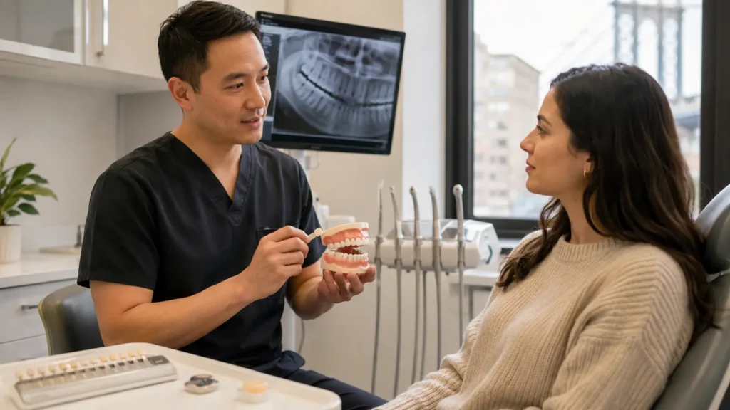
Restorative Dentistry Guide · Williamsburg Brooklyn Do you really need a metallic-looking filling — or is there an option that blends in and still does the job? For most small to moderate cavities, the answer is a composite resin filling. Tooth-Colored Fillings in Brooklyn: Natural-Looking Cavity Repair COMPOSITE VS. SILVER WHAT TO EXPECT Written by Toothology Editorial Team | Reviewed by Dr. Ayala Lieberman, DDS June 2026 Hearing “cavity” at a routine checkup is never ideal. Or maybe the concern isn’t new — you’ve had a silver filling in a back molar since you were a teenager, and now you’re wondering whether it’s time for something more natural-looking. Either way, many patients ask the same question: do I really need a metallic-looking filling, or is there an option that blends in and still does the job? Tooth-colored fillings are often the answer. For many small to moderate cavities, a composite resin filling can repair decay, restore function, and match your natural tooth colour closely enough that most people won’t notice it. What Are Tooth-Colored Fillings? A tooth-colored filling — also called a composite filling or composite resin filling — is a dental restoration made from resin mixed with fine glass or ceramic particles. Unlike silver amalgam fillings, composite resin is shaded to match your natural tooth colour, so the repair blends in naturally. Bonds Directly to Tooth Composite adheres to the tooth with a dental adhesive. In many cases this allows the dentist to remove less healthy tooth structure compared with traditional fillings. Front and Back Teeth Tooth-colored fillings can be used on both front and back teeth. They are especially useful for visible front teeth, and commonly used for small to moderate cavities in back teeth. Shade-Matched to You The dentist selects the closest composite shade to your natural enamel before placing the filling, so the final result blends rather than contrasts. Mercury-Free Material Composite resin contains no mercury — a common preference for patients who want an alternative to silver amalgam for new fillings or replacements. NOT RIGHT FOR EVERY TOOTH For larger cavities or heavily damaged teeth, your dentist may recommend a different restoration — such as a crown or onlay — rather than a composite filling. The recommendation depends on how much healthy tooth structure remains. How Composite Fillings Repair Cavities The dental filling procedure for a composite resin filling is usually simple and completed in one visit. Knowing the steps makes the appointment feel more routine and less stressful. 1 DECAY REMOVAL The dentist removes the decayed or weakened part of the tooth, leaving healthy structure behind. Because composite bonds to the tooth, this is often more conservative than preparing for a silver filling. 2 BONDING A dental adhesive is applied so the composite resin can attach securely to the tooth’s surface. 3 LAYERING The composite material is placed in thin layers to rebuild the tooth shape accurately and ensure a strong, seamless result. 4 CURING Each layer is hardened with a special curing light — the process takes only seconds per layer. 5 SHAPING & BITE CHECK The filling is shaped to match the natural contours of your tooth and bite. Your dentist checks that it doesn’t sit too high or cause discomfort when biting. 6 POLISHING The surface is polished so it looks smooth and natural, and is less likely to collect plaque or stain over time. ONE APPOINTMENT, SAME DAY For a straightforward cavity, the process is typically calm, routine, and finished in a single appointment. Most patients return to normal daily activities the same day, once any numbness has worn off. Tooth-Colored Fillings vs. Silver Fillings Both tooth-colored and silver fillings have been used successfully in dentistry. The right choice depends on the tooth, the size of the cavity, your cosmetic goals, and your dentist’s recommendation. Factor Tooth-Colored (Composite) Silver (Amalgam) Appearance Matches natural tooth shade; blends with surrounding teeth Metallic grey/silver; visible, particularly on front or easily seen back teeth Material Composite resin — resin with fine glass or ceramic particles Amalgam — a metal alloy including mercury, silver, tin, and copper Bonding Bonds directly to tooth structure with a dental adhesive Held in place more by mechanical retention within the prepared cavity Best Fit Small to moderate cavities, visible areas, cosmetic preference Case dependent; many existing amalgam fillings remain functional for years Patient Priority Usually Driving the Choice Natural-looking result; mercury-free material preference Durability track record; less concern about visible colour A BALANCED WORD ON EXISTING SILVER FILLINGS Many older silver fillings still work well and do not need replacement just because they are old or visible. If a silver filling is intact, comfortable, and free of decay, replacement may not be necessary. However, if it is cracked, leaking, has decay around it, or you prefer a more natural-looking material, replacing it with composite can be a reasonable option — worth discussing at your next checkup. When Is a Filling Enough to Save a Tooth? Composite fillings are a conservative option, but they’re not right for every tooth. A filling usually works best when the cavity or damage is small to moderate and there is enough healthy tooth structure left to support the repair. A FILLING USUALLY WORKS WHEN: The cavity is small to moderate Enough healthy tooth structure remains to support the repair No significant crack or fracture is present The tooth has not had multiple previous fillings leaving weakened walls A CROWN MAY BE NEEDED WHEN: The cavity is large and has removed too much tooth structure A crack extends into the tooth A cusp is broken The tooth has had multiple fillings and little strong structure remains Root canal treatment has been completed on a back tooth ONLY AN EXAM CONFIRMS WHICH TREATMENT IS RIGHT The only way to know what your tooth needs is through an exam and X-rays. Your dentist will check the damage, the remaining tooth structure, and the bite pressure before recommending a filling, crown, or another restoration. What Happens During a Dental Filling Appointment?
Porcelain Crowns: When a Damaged Tooth Needs More Than a Filling
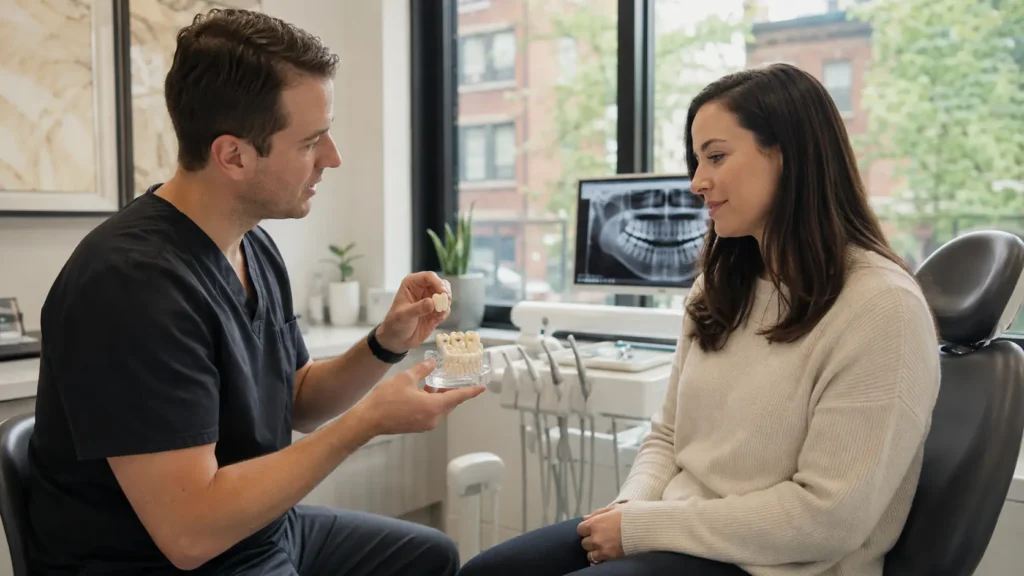
Restorative Dentistry Guide · Williamsburg Brooklyn A sharp twinge that hasn’t gone away. An old filling finally failing. A crown that’s come loose. The same question keeps coming up: is a filling enough, or do you need a crown? Porcelain Crowns: When a Damaged Tooth Needs More Than a Filling FILLING VS. CROWN SEE THE WARNING SIGNS Written by Toothology Editorial Team | Reviewed by Dr. Ayala Lieberman, DDS June 2026 You bit into something ordinary — bread, an apple, nothing dramatic — and felt a sharp twinge that hasn’t fully gone away. Or maybe an old, dark filling your dentist has been watching for years is finally starting to fail. Or perhaps a crown came loose, and you’re wondering whether it can simply be reattached. In all of these situations, the same question comes up: is a filling enough, or do I need a porcelain crown? A porcelain crown is a custom-made cap that covers and protects the visible part of a damaged tooth. Sometimes it’s the right choice; other times, a filling, bonding, or smaller restoration may be enough. The answer depends on how much healthy tooth remains, where the damage is, and how much chewing force the tooth needs to handle. IF A CROWN HAS COME LOOSE Do not try to glue it back yourself. In some cases, a dentist can re-cement the crown — but only after checking whether it still fits properly and whether the tooth underneath has decay, fracture, or other damage. What Is a Porcelain Crown? A porcelain crown is a custom-made cap that fits over the visible part of a damaged or weakened tooth. It is shaped and shaded to look natural while protecting the tooth underneath. F A FILLING REPAIRS PART OF A TOOTH If the damage is small, a filling can fully restore the affected portion while leaving the rest of the natural tooth structure intact. C A CROWN PROTECTS THE WHOLE TOOTH When a tooth has a large cavity, crack, broken cusp, or multiple old fillings, the remaining structure may be too weak to support another filling — full-coverage protection becomes the safer option. The recommendation is based on the tooth’s condition — not just appearance. When Is a Crown Better Than a Filling? The choice between a filling and a crown usually comes down to one question: is there enough healthy tooth structure left to support a filling under normal chewing force? If the tooth walls are too thin, cracked, or weakened, a crown may be the safer long-term option. Large Cavity If decay has removed too much tooth structure, a filling may not be strong enough. A crown can protect the remaining tooth from cracking under bite force. Cracked Tooth When a crack extends into the tooth, a crown can help hold the tooth together and reduce stress during chewing. Broken Cusp If one of the raised chewing points breaks, the tooth may need a crown to rebuild strength and restore normal function. Failing Old Filling Repeated filling replacements remove more natural tooth each time. When too little strong structure remains, a crown is often more durable. After Root Canal Treatment Many back teeth need crowns after root canals because they can become weaker and more prone to fracture. Your dentist will decide based on how much tooth remains and how much bite force it handles. Filling vs. Crown: A Quick Comparison Factor Filling Porcelain Crown Best For Small to moderate cavities with enough healthy tooth left Large cavities, cracks, broken cusps, or weakened teeth Protection Level Replaces the damaged portion only Covers and protects the whole visible tooth Longevity Works well for smaller repairs Often stronger for heavily damaged teeth Invasiveness More conservative Requires more shaping of the tooth When It Falls Short Large fillings can weaken or fracture over time Not needed for minor damage CHOOSING THE WRONG TREATMENT COSTS MORE LATER Trying to use a filling when a tooth really needs a crown can lead to cracks, breakage, or a future dental emergency. Choosing the right treatment early often helps protect the tooth and avoid a more complex repair down the line. Signs You May Need a Dental Crown While only an exam and X-rays can confirm whether a tooth needs a crown, there are recognisable signs and situations that warrant scheduling that evaluation rather than waiting. Visible crack lines on the tooth surface Pain or a sharp sensation specifically when biting down A large, old filling that covers much of the tooth A fractured or broken cusp — the pointed part of a back tooth Recurring decay around or beneath an existing filling A tooth that has been significantly worn down over time An old crown that is cracked, loose, or visibly damaged A tooth that has had root canal treatment and has not yet been crowned Warning Signs That Need Prompt Evaluation Sharp pain specifically when biting down Swelling near a tooth Worsening sensitivity to hot, cold, or pressure A visibly broken tooth edge A crown that has fallen off entirely THESE SIGNS DON’T CONFIRM A CROWN — BUT THEY CONFIRM AN EXAM These signs do not automatically mean you need a crown, but they do mean an exam and X-rays are the right next step. Your dentist can then decide whether a filling, crown, or another treatment is most appropriate for your specific tooth. Porcelain Crowns vs. Ceramic Crowns Patients often use porcelain and ceramic interchangeably, and that’s usually fine. Technically, porcelain is a type of ceramic material, often chosen for its natural tooth-like colour and translucency. Zirconia crowns are also ceramic, but they’re denser and known for strength — making them useful for back teeth that handle heavier chewing forces. Some cases may also use porcelain-fused-to-metal crowns, though all-ceramic and zirconia options are now more common. All-Ceramic / Porcelain vs. Zirconia: A Quick Comparison Factor All-Ceramic / Porcelain Zirconia Aesthetics Excellent shade-matching and translucency; often preferred for visible front teeth Good aesthetics with newer materials, but sometimes slightly less translucent
Emergency Root Canal in Brooklyn: Signs You Need Same-Day Treatment

Emergency Dental · Same-Day Root Canal · Brooklyn The pain has been building since last night — throbbing, constant, relentless. Here’s how to tell if you need same-day root canal treatment, what the procedure involves, and what it costs in Brooklyn. Emergency Root Canal in Brooklyn: Signs You Need Same-Day Treatment CALL US NOW SEE THE WARNING SIGNS Written by Toothology Editorial Team | Reviewed by Dr. Ayala Lieberman, DDS June 2026 The pain has been building since last night. It’s throbbing now — not a dull ache that comes and goes, but a constant, relentless pressure that’s making it impossible to focus, eat, or sleep. You’ve taken whatever pain relief you had on hand and it barely touched it. If this is your situation, this article is for you. An emergency root canal in Brooklyn is not the right answer for every bad toothache — but for certain tooth pain symptoms, it is the correct and urgent response. Recognising the difference between ordinary sensitivity and pulp inflammation or dental abscess helps you make the right call today rather than in another day or two of worsening pain. Signs Your Toothache May Require an Emergency Root Canal Not every toothache is a same-day emergency. Brief cold sensitivity or mild sensitivity after recent dental work can come from exposed dentin rather than an infected nerve. But the following signs are more concerning and should be evaluated promptly — the same day if possible. Throbbing pain without a trigger — pain that starts on its own, especially worsening at night, can suggest pulp inflammation or infection Pain that wakes you up — tooth pain that interrupts sleep is a strong sign the nerve may be seriously inflamed Heat sensitivity that lingers — brief cold sensitivity is common, but heat pain that stays after the trigger is removed is more concerning for pulp involvement Severe pain when biting or touching the tooth — this can mean infection or inflammation has reached the root area and surrounding tissue Localised swelling or a pimple-like bump on the gum — this may signal an abscess draining near the root tip Persistent bad taste or bad odour from one area — this can occur when infection is actively draining WHY LINGERING HEAT PAIN IS MORE CONCERNING THAN COLD SENSITIVITY Brief cold sensitivity that goes away quickly is often caused by exposed dentin. Lingering heat pain is more concerning because it can suggest the nerve inside the tooth is irreversibly inflamed. In those cases, the tooth may not recover on its own and may need root canal treatment. When to Go to the ER or Call 911 Instead Swelling spreading to the throat, floor of the mouth, or neck Difficulty breathing, swallowing, or speaking Swelling under the eye or rapidly worsening facial swelling High fever combined with dental swelling Tightness or pressure in the throat Swelling that is spreading quickly — over hours rather than days For ER-Level Symptoms — Don’t Call a Dentist First Spreading swelling toward the jaw, neck, or throat, and any difficulty breathing or swallowing, is a medical emergency. Go to the ER or call 911 immediately. For severe tooth pain, localised swelling, bad taste, or worsening symptoms over 24–48 hours, call Toothology for a same-day urgent appointment. What Happens During a Same-Day Endodontic Procedure? The term “root canal” still scares many people — but with modern local anaesthesia, most patients experience it as pressure and movement rather than sharp pain. Knowing what usually happens makes the visit far less intimidating than the idea of it. 1 URGENT EXAM & X-RAYS The dentist checks the tooth and takes imaging to confirm the source of pain, the extent of infection, and whether the tooth can still be saved. 2 LOCAL ANAESTHESIA Local anaesthetic fully numbs the tooth and surrounding area. Extra anaesthesia may be needed if inflammation is making the tooth harder to numb — a known challenge that experienced clinicians manage with targeted techniques. 3 ACCESS OPENING A small opening is made through the top of the tooth to reach the pulp chamber where the infected tissue is located. 4 PULP REMOVAL & CANAL SHAPING The inflamed or infected pulp is carefully removed from the pulp chamber and root canals, which are then cleaned and shaped to prepare for sealing. 5 IRRIGATION & DISINFECTION The inside of the tooth is flushed with irrigating solution to remove bacteria and debris from the root canal system. 6 SEALING THE CANALS In some cases the canals are sealed the same day; in others, a temporary medicated filling is placed first and the seal completed at a second visit depending on infection severity. 7 TEMPORARY OR PERMANENT CLOSURE + CROWN The tooth is closed with a temporary or permanent material. A dental crown is typically recommended afterward to protect the tooth long-term — especially for molars and premolars. CAN A THROBBING TOOTH STILL BE SAVED? Often yes. Severe pain does not automatically mean the tooth is beyond saving. In many cases, pain comes from pressure and infection inside the tooth, and once that infected pulp is removed, the pain improves significantly within a day or two. Whether it can be saved depends on how much healthy tooth structure remains — determined by exam and X-rays. WHAT HAPPENS IF YOU DELAY A ROOT CANAL? An infected pulp does not heal on its own. Without treatment, the infection can spread into surrounding bone, create an abscess, and in more serious cases spread into deeper facial or neck spaces. Even before that, waiting can mean more bone loss, more difficult treatment, and a tooth that becomes harder or impossible to save. How Local Anaesthesia Numbs the Area During Root Canal Treatment Fear of pain during a root canal is one of the main reasons people delay treatment. In reality, most patients find the procedure itself far more manageable than the
Lost a Filling? What to Do Before Seeing an Emergency Dentist

Urgent Dental Guide · Williamsburg Brooklyn Something shifted while eating — your tooth feels rough, hollow, or sharply different. Here’s exactly what to do right now, when it’s a same-day emergency, and what to expect at the repair visit. Lost a Filling? What to Do Before Seeing an Emergency Dentist CALL US NOW 8-STEP ACTION PLAN Written by Toothology Editorial Team | Reviewed by Dr. Ayala Lieberman, DDS June 2026 You’re eating, something hard shifts in your mouth, and suddenly a tooth feels rough, hollow, or sharply different than it did a moment ago. Sometimes the filling comes out in one piece. Sometimes there’s a sharp edge or sudden sensitivity. Either way, the question is the same: how serious is this, and what do you do right now? A lost filling can range from uncomfortable to genuinely urgent depending on your symptoms. Mild sensitivity may be manageable until the next urgent appointment with some short-term protection. But biting pain, throbbing, swelling, or a larger fracture should not wait. Whatever the severity, a missing filling should be checked promptly — the tooth underneath is now exposed, and what starts as manageable sensitivity can become a bigger problem if decay, a crack, or nerve involvement is left untreated. What to Do Immediately If Your Dental Filling Falls Out The first priority is to protect the exposed tooth and keep it comfortable until you can be seen. Rinse gently with warm water to clear away debris without disturbing the area or surrounding tissue. Save the filling if you find it and bring it to your appointment — it helps the dentist understand what came out and why. Do not use household glue or any adhesive to try to stick it back in — they’re not designed for oral use and can complicate treatment. Avoid chewing on that side to prevent additional damage, fracture, or further loss of tooth structure. Avoid very hot, cold, or sweet foods and drinks that trigger sensitivity — these stimulate the exposed dentin directly. Use over-the-counter pain relief if needed and only as directed on the label or by your pharmacist. A pharmacy temporary filling kit may help short-term — but it is only a bridge measure. It does not repair the tooth and should not delay seeking dental care. Call a dentist promptly. A lost filling will not fix itself, and the exposed tooth needs professional evaluation — not a wait-and-see approach. When a Lost Filling Becomes a Same-Day Dental Problem Not every lost filling is a same-day emergency — but some are. The pattern of your symptoms is the key indicator. What You Notice What It May Suggest Urgency What to Do Rough or hollow feeling; mild sensitivity to cold or air; no pain at rest Filling lost, dentin exposed — manageable for a short window with temporary protection in place Next Available Use a temporary filling kit as a bridge; call for the next available urgent appointment — do not delay beyond a few days Cold or heat sensitivity that lingers more than a few seconds after the trigger is removed More significant dentin exposure; possibly early pulp involvement; needs evaluation to rule out nerve involvement Same/ Next Day Book a same-day or next-day urgent appointment; avoid further temperature triggers until seen Sharp pain when biting or releasing bite pressure Possible crack extending into the tooth — the old filling may have been holding the tooth together under load Same Day Book a same-day urgent appointment; avoid biting on that side entirely until evaluated by a dentist Spontaneous throbbing at rest, bad taste, swelling of gum or cheek, or fever Advanced infection, abscess, or significant structural failure — requires immediate professional attention Today — Urgent Call the practice immediately or visit urgent dental care today; go to the ER if swelling spreads toward the jaw, neck, or throat Go to the ER immediately if swelling is spreading toward the jaw, neck, or throat — or if you have any difficulty breathing or swallowing. This is a medical emergency, not a dental appointment situation. Go to the ER Immediately If: Swelling is spreading to the jaw, throat, or neck — or if you have difficulty breathing or swallowing. This is a medical emergency, not a dental appointment situation. Why a Missing Filling Causes Sharp Hot and Cold Sensitivity A filling seals and protects the inner part of the tooth. When it falls out, the dentin beneath becomes exposed. Dentin contains tiny microscopic channels — dentinal tubules — that connect toward the nerve. That’s why cold water, hot drinks, sweets, or even air can suddenly cause sharp sensitivity that wasn’t there before. COLD VS HEAT SENSITIVITY Cold is often the strongest trigger, but hot foods and sweets can also cause discomfort depending on how much tooth is exposed and how close the area is to the nerve. Sensitivity to heat in particular can suggest the nerve is more significantly involved and warrants same-day evaluation. Why Biting Pain After a Filling Falls Out Matters More Sensitivity to temperature is common after a filling falls out — but pain when biting is more concerning. That kind of pain can suggest the tooth is cracked. In some cases, the old filling was helping hold part of the tooth together. Once it’s gone, the cracked area can move slightly under biting pressure and irritate the nerve. Biting pain after a lost filling should not be managed with OTC medication alone — it should be evaluated the same day or next morning. What Happens If the Tooth Underneath Is Cracked A smaller crack may be repairable with a new filling or a crown A deeper crack may require a root canal before the final restoration If the crack extends too far below the gumline , the tooth may not be restorable THE SOONER A CRACK IS FOUND, THE MORE OPTIONS THERE ARE A crack identified early may be a simple filling or crown. The same crack identified weeks later may require extraction.
Is It Better to Get a Dental Bridge or an Implant? A Patient’s Guide

Restorative Dentistry Guide · Williamsburg Brooklyn Both options replace a missing tooth. Both can look and feel natural. But they work in completely different ways — and the differences affect your neighboring teeth, your jawbone, your timeline, and what you’ll actually pay over time. Dental Bridge vs. Implant: A Patient’s Guide SEE THE FULL COMPARISON QUESTIONS TO ASK YOUR DENTIST Written by Toothology Editorial Team | Reviewed by Dr. Ayala Lieberman, DDS June 2026 One dentist mentions a bridge. Another mentions an implant. A family member had one experience; a colleague had another. And somewhere in the middle of all that input, you are sitting with a missing tooth — or an extraction coming up — trying to make a decision that involves surgery, cost, timeline, and something called osseointegration. The answer genuinely depends on your individual circumstances. Both options can effectively replace a missing tooth. Both can restore the appearance, function, and confidence that come with a complete smile. But they solve the same problem in very different ways — and those differences have direct implications for your neighboring teeth, your jawbone, your long-term treatment plan, and what you will actually pay over time, not just upfront. Replacing a Missing Tooth: Bridges vs. Implants Both a bridge and an implant can replace a missing molar, restore chewing, and close the space. The main difference is how they are supported. A fixed dental bridge uses the teeth on either side of the gap as anchors. Those neighboring teeth are usually shaped for crowns, and the artificial tooth is attached between them as one cemented unit. A single tooth implant replaces the missing root with a titanium post placed in the jawbone. After healing, a crown is attached on top — so the new tooth functions independently of the teeth next to it. FIXED DENTAL BRIDGE Faster · No surgery · Relies on adjacent teeth Uses neighboring teeth as anchors — those teeth are shaped down for crowns Artificial tooth is cemented between them as one fixed unit No surgery — conventional dental work only Completed in 2–3 visits over a few weeks Generally lower upfront cost Does not replace the missing root in the bone SINGLE TOOTH IMPLANT Longer timeline · Surgical · Stands independently Titanium post placed in the jawbone — replaces the missing root Crown attached on top after healing — functions independently Surgical placement under local anaesthesia Several months total — accounts for osseointegration Higher upfront cost; potentially lower total cost over time Helps preserve bone at the missing-tooth site Side-by-Side: The Key Differences at a Glance Factor Fixed Dental Bridge Single Tooth Implant Upfront Cost Generally lower; bridge + abutment crowns completed in one treatment phase Higher; accounts for surgery, implant fixture, healing period, and final crown Treatment Timeline Typically 2–3 appointments over 2–4 weeks total Several months; accounts for osseointegration and possible bone grafting Surgery Required No surgery — conventional dental work only Yes — surgical placement of titanium post under local anaesthesia Effect on Neighboring Teeth Adjacent teeth prepared as abutments; involves reshaping healthy tooth structure if not already crowned No impact on neighboring teeth — stands completely alone in the jaw Bone at Missing-Tooth Site Not directly loaded; some bone resorption at the edentulous site may occur gradually over time Implant post stimulates the bone it integrates with, helping preserve bone volume at the site Maintenance Requires floss threaders or interdental aids to clean under the bridge — cannot be flossed conventionally Brushed and flossed like a natural tooth; professional monitoring of peri-implant tissue at cleanings Long-Term Replacement Risk Abutment teeth can develop problems over time; bridge may eventually need replacing as a complete unit Implant post designed to be long-lasting; crown on top may eventually need replacement independently Neither option is universally better. The right choice depends on bone support, the condition of neighboring teeth, budget, timeline, and long-term goals. A case-specific evaluation determines which fits your situation. The Surgical Realities of a Titanium Implant Post An implant is a surgical treatment — and that matters in the decision. It involves anaesthesia, healing time, and a longer overall process than a bridge. For many patients, the long-term benefits make that worthwhile. For others — those with limited bone, medical concerns, tight timelines, or strong hesitation about surgery — a bridge may be the more practical option. How Long Each Option Actually Takes A fixed dental bridge is usually completed in two to three visits over a few weeks. The neighboring teeth are prepared, a temporary is often placed while the lab fabricates the bridge, and the final unit is cemented once ready. A single tooth implant usually takes several months, depending on healing and whether extra procedures are needed first. 1 CONSULTATION & 3D IMAGING Exam and imaging to assess bone volume, nearby anatomy, and implant suitability. CBCT 3D imaging is often used for precise surgical planning. 2 BONE GRAFTING (IF NEEDED) Some patients need grafting first if bone has been lost at the site — either from the extraction or time elapsed. This adds time and cost but makes implant placement viable in cases that otherwise wouldn’t qualify. 3 IMPLANT SURGERY The titanium post is surgically placed into the jawbone under local anaesthesia. Sedation options are available for anxious patients. 4 OSSEOINTEGRATION — HEALING PHASE The bone gradually bonds to the implant over several months. This is the longest phase — and it cannot be rushed. Regular monitoring confirms integration is progressing correctly. 5 ABUTMENT & FINAL CROWN Once healing is confirmed, the abutment connector and final custom crown are attached. The result is a fully functional, natural-looking tooth. MANAGING THE GAP WHILE HEALING If timing or appearance during the healing phase is a concern, temporary options can often be discussed. Ask about temporary solutions at your consultation — leaving a visible gap for several months is not usually necessary. Why a Bridge Requires Altering Healthy Neighboring Teeth This is often the factor that pushes the decision toward an implant when the teeth next to the gap are still healthy and
All-on-4 Dental Implants vs. Dentures: Which Is the Better Investment?

Full-Arch Restoration Guide · Williamsburg Brooklyn An honest comparison — stability, bone health, cost of ownership over 10 years, and who is the right candidate for each option. No overselling either direction. All-on-4 Dental Implants vs. Dentures: Which Is the Better Investment? SEE THE FULL COMPARISON WHO IS THE RIGHT CANDIDATE? Written by Toothology Editorial Team | Reviewed by Dr. Ayala Lieberman, DDS April 2026 If you are living with dentures — or facing the prospect of significant tooth loss — you have almost certainly heard the term “All-on-4 dental implants” come up in conversation, in advertising, or in a consultation room. The honest answer is that dentures vs. implants is not a simple better/worse question. It depends on your bone health, medical history, financial situation, and what you are actually hoping to change about your current experience. Neither option should be approached as a one-size-fits-all decision. Dentures can be the right solution for many patients — particularly where surgical contraindications exist or where cost is the overriding constraint. All-on-4 is a surgical procedure with a recovery process and specific candidacy requirements. What follows is an honest account of both. The Difference Between Removable Dentures and All-on-4 Implants The most fundamental distinction between the two options is fixed versus removable. Removable dentures sit on top of the gum tissue and are held in place by suction, clasps on remaining teeth, or dental adhesive. They come out at night, require soaking and cleaning in a separate container, and rely on the continued fit of the gum tissue beneath them — which changes as the underlying bone resorbs over time. All-on-4 is a full-arch implant concept in which the entire replacement arch of teeth is supported by four implants placed strategically in the jawbone. The prosthetic — often a zirconia bridge or acrylic hybrid on a titanium framework — is fixed to the implants and does not come out for daily cleaning. It does not require adhesive, does not rely on gum suction, and because the implants are embedded in bone, they stimulate it in a way that removable dentures do not. NOTE ON “PERMANENT DENTURES” This term is used inconsistently — it sometimes refers to dentures that are implant-supported and non-removable, and sometimes to high-quality conventional dentures intended for long-term use. A fixed implant-supported arch is more accurately described as a full-arch implant bridge rather than a denture in the conventional sense. Full Side-by-Side Comparison Category Removable Dentures All-on-4 Implants Daily Stability Relies on suction, adhesive, or clasps; can shift during speaking and eating Fixed to implants; does not move, slip, or require adhesives during the day Bite Force Significantly reduced compared to natural teeth; hard foods often avoided Substantially stronger than removable dentures; allows a wider range of foods (case-dependent) Jawbone Preservation Does not stimulate bone; continued resorption changes face shape and denture fit over time Implants stimulate bone similarly to natural tooth roots, helping to slow ongoing resorption Daily Care Removed nightly, soaked, brushed; adhesive reapplied each morning Cleaned in place with soft brush, floss tools, water flosser; removed only by clinician Adjustments & Repairs Periodic relines needed as bone changes; repairs for cracks or fractures; replacement over time Periodic check-ups; prosthetic may need adjustment or replacement over many years; implants typically remain Comfort Over Time Fit can worsen as bone shrinks; sore spots and gum irritation are common complaints No gum pressure from a removable plate; discomfort during healing period is temporary Speech Initial adjustment period; clicking or slipping can affect speech, particularly without adhesive Fixed prosthetic closely mimics natural tooth position; most patients adapt quickly Long-Term Appearance Facial appearance may change over time as underlying bone is resorbed Implants help preserve bone contour, supporting a more stable facial profile over time Why All-on-4 Is Often Called “Teeth in a Day” The phrase “Teeth in a Day” — sometimes described as same-day implants or immediate loading — refers to the possibility of placing implants and attaching a temporary fixed prosthetic within a single surgical appointment. The concept involves strategically angling implants — often tilting the posterior implants to avoid the sinus cavity and areas of reduced bone density — to maximise contact with available bone. WHAT “SAME DAY” ACTUALLY MEANS “Same day” refers to the temporary bridge, not the final bridge. The final prosthetic comes after osseointegration is confirmed — typically several months later. And not every patient qualifies for immediate loading — this is determined at consultation with 3D imaging. What the Full Treatment Process Typically Involves 1 CONSULTATION & 3D IMAGING A thorough examination including CBCT (3D cone beam CT) imaging to assess bone volume, bone density, nerve positions, sinus anatomy, and overall jaw suitability for implant placement. 2 TREATMENT PLANNING Reviewing options for number of implants, prosthetic materials (zirconia bridge, acrylic hybrid), timeline, and any prerequisite treatment needed — including extractions or bone grafting. 3 EXTRACTIONS (IF NEEDED) Remaining failing teeth are extracted — often on the same day as implant placement in qualifying cases, reducing the total number of surgical appointments. 4 IMPLANT PLACEMENT Implants are surgically placed into the jawbone under local anaesthesia, with sedation options available. Implants are tested for primary stability before any immediate loading decision is made. 5 TEMPORARY FIXED BRIDGE (SAME DAY — IF QUALIFIED) In cases where sufficient primary stability is achieved, a temporary prosthetic is attached to the implants on the same day — allowing you to leave with fixed, functional teeth. 6 HEALING & OSSEOINTEGRATION Over several months, the implants fuse with the surrounding bone. Regular monitoring appointments assess integration progress and ensure healing is on track. 7 FINAL PROSTHETIC Once osseointegration is confirmed, the temporary bridge is replaced with the final restoration — often a full-arch zirconia bridge or similar durable prosthetic designed for long-term wear. Long-Term Benefits: Bone Health and Dietary Freedom Jawbone Preservation: Why It Affects Your Face, Not Just Your Teeth When natural teeth are lost, the bone that previously surrounded their roots no longer receives the stimulation it needs to maintain its density and volume. Without that stimulation,
Why Are My Teeth Suddenly Sensitive to Cold Water? Causes and Treatment

Tooth Sensitivity Guide · Williamsburg Brooklyn That sharp zing when you sip cold water isn’t just annoying — it’s a signal. Cold sensitivity is a symptom, not a diagnosis. Here’s what’s actually behind it and what fixes it. Why Are My Teeth Suddenly Sensitive to Cold Water? Causes & Treatment FIND THE CAUSE SEE THE TREATMENTS Written by Toothology Editorial Team | Reviewed by Dr. Ayala Lieberman, DDS June 2026 You take a sip of iced coffee and feel a sharp zing in your tooth. Cold air hits your teeth on a winter morning and you wince. A glass of cold water suddenly feels different than it did a few months ago. Cold sensitivity is one of the most common dental complaints — but also one of the most misunderstood. Some people assume it’s normal and reach for a desensitising toothpaste. Sometimes that helps. Other times, it only masks a bigger issue like a cavity, crack, gum recession, or failing filling. The key thing to understand is that cold sensitivity is a symptom, not a diagnosis. Mild sensitivity across several teeth is different from sharp, lingering pain in one tooth — and those differences matter when deciding what to do next. What Triggers Sharp, Sudden Temperature Sensitivity? When cold water causes a sharp zing, the problem is usually not the enamel itself. Enamel is the hard outer layer of the tooth and does not contain nerves. Beneath it is dentin, which contains tiny channels called dentinal tubules that connect toward the inner nerve of the tooth. When dentin becomes exposed — from enamel wear, gum recession, a crack, or a cavity — cold water or air can trigger movement inside those tubules. That movement stimulates the nerve and creates the quick, sharp pain people describe as tooth sensitivity. UNDERSTANDING TOOTH LAYERS — WHY COLD REACHES THE NERVE ENAMEL — OUTER SHELL The hardest substance in the body. Contains no nerves. When intact, it fully insulates the dentin and pulp. When worn or eroded, dentin sits closer to the surface. DENTIN — THE SENSITIVE LAYER Contains dentinal tubules — microscopic channels running toward the nerve. Exposed dentin is the direct cause of cold sensitivity. Root surfaces have no enamel — only thinner cementum — making them especially reactive. PULP & NERVE — INNER CORE Soft tissue containing the tooth’s nerve supply. Pain that lingers several seconds after the cold trigger is gone may suggest the nerve is significantly involved. WHY COLD WATER HURTS MORE THAN ROOM-TEMPERATURE WATER Cold water triggers a stronger response because it causes a faster fluid shift inside exposed dentinal tubules. That sudden movement stimulates the nerve and produces the sharp, immediate pain people notice. The same response can happen with cold air — particularly when root surfaces or worn enamel are exposed. Acidic foods and drinks can make this temporarily worse by softening the surface and opening tubules further. WHY “SUDDEN” SENSITIVITY MATTERS When sensitivity feels sudden or is noticeably worsening, it often means something has changed. Gum recession may have exposed more root surface. Enamel may have worn further. A crack may have formed. Or a filling may no longer be sealing the tooth properly. Sensitivity that appears recently or escalates quickly deserves more attention than sensitivity that has stayed mild and stable over a long period. How Gum Recession Uncovers Tooth Nerve Paths Enamel only covers the crown of the tooth — the visible portion above the gumline. Below the gumline, the root surface is covered by cementum: a thinner, far less protective material. When gum tissue recedes — pulling away from the tooth and exposing part of the root — that exposed surface produces a much more pronounced sensitivity response than comparable dentin exposure on the crown. Gum recession is one of the most common causes of cold sensitivity. It often develops slowly — which is why the pain can feel sudden once enough root surface finally becomes exposed. Aggressive Brushing Brushing too hard or using a medium or hard-bristle toothbrush can gradually wear the gumline and expose sensitive root surfaces over months to years. Abrasive Toothpaste Some whitening or “heavy-cleaning” toothpastes can increase gumline wear — particularly when combined with forceful brushing technique. Periodontal Disease Gum disease causes the gums and supporting bone to pull away from the tooth, leading to more significant recession — often across multiple teeth simultaneously. Naturally Thin Gum Tissue Some people naturally have thinner gum tissue, which makes recession more likely over time regardless of hygiene habits. PRODUCT CHANGES AREN’T ENOUGH Switching to a softer toothbrush and less abrasive toothpaste can help prevent further damage — but it won’t restore gum tissue that has already receded, address gum disease if that’s the cause, or tell you how severe the recession is or whether it’s still progressing. A dental evaluation is more useful than relying on a product change alone. In-Office Desensitising vs. Store-Bought Sensitivity Toothpaste Sensitive teeth treatments range from over-the-counter toothpaste to in-office desensitising procedures. The right option depends on the cause and severity of the sensitivity. When Sensitivity Toothpaste Is a Reasonable Starting Point Sensitivity toothpastes with ingredients like potassium nitrate or stannous fluoride can help reduce mild, generalised sensitivity by calming the nerve response or blocking exposed dentinal tubules. They can be a reasonable first step for minor enamel wear or temporary post-whitening sensitivity. THE LIMITATION OF TOOTHPASTE ALONE Toothpaste only helps manage the symptom. It does not treat the underlying cause if the problem is gum recession, a crack, a failing filling, or early decay. If sensitivity is worsening, localised, or persistent — relying on toothpaste alone can delay the right diagnosis and allow the underlying issue to worsen. How Treatment Options Compare Treatment Option What It Does Best Suited For At-Home Sensitivity Toothpaste Potassium nitrate / stannous fluoride Calms nerve endings in dentin tubules over repeated use; gradual improvement over several weeks of consistent use Mild diffuse dentin hypersensitivity; ongoing maintenance for sensitivity-prone patients; first step for minor enamel wear In-Office Fluoride Treatment or Varnish Concentrated mineral application
Wisdom Teeth Removal in Williamsburg: How 3D Imaging Makes Surgery Safer
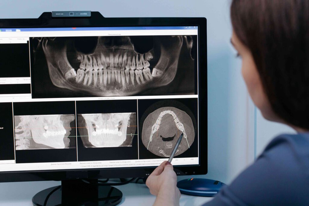
Wisdom Teeth Removal Guide · Williamsburg · 3D Imaging CBCT 3D imaging shows the exact path of the nerve, root shape, and jaw anatomy that a flat 2D X-ray can’t. Here’s why it changes the surgical plan — and what recovery actually looks like. Wisdom Teeth Removal in Williamsburg: How 3D Imaging Makes Surgery Safer HOW 3D IMAGING HELPS THE 48-HOUR RECOVERY PLAN Written by Toothology Editorial Team | Reviewed by Dr. Ayala Lieberman, DDS June 2026 For most people, wisdom teeth removal is something they know is coming long before it becomes urgent. A dentist mentions it at a routine checkup. An X-ray shows third molars heading in the wrong direction. Or the jaw starts aching in a way that feels different from anything before. Then comes the question most people delay longer than they should: when is the right time, and how do I make sure this goes as smoothly as possible? Wisdom teeth removal is one of the most commonly performed dental procedures, and for the vast majority of patients it goes without significant complication. But the cases that generate the most anxiety, and the most serious post-operative concerns, often involve anatomy that isn’t fully characterised on initial 2D imaging — especially when roots appear close to important nerves. Modern CBCT 3D imaging has changed that picture significantly. Why 3D Nerve Mapping Is Essential for Safe Third Molar Extraction The inferior alveolar nerve — which supplies sensation to the lower lip, chin, and lower teeth — runs in a canal in the lower jaw near the roots of lower wisdom teeth. In simple, fully erupted cases, a panoramic X-ray may be enough. But with deep impactions or roots that appear close to the nerve on a 2D image, there can be real uncertainty. A CBCT (cone beam CT) 3D scan gives a full three-dimensional view of the jaw so the surgeon can see the exact relationship between the roots and the nerve canal — information that can directly affect the surgical plan. Whether to Section the Tooth Removing the tooth in smaller pieces can reduce force near nerve-adjacent roots. CBCT helps guide that decision more precisely than a flat image can. Which Approach to Use 3D mapping can influence the entry angle, how much bone needs to be removed, and the safest direction of controlled pressure during extraction. Whether to Refer CBCT may reveal complexity that is better handled by a specialist — far better to know before surgery than to discover it mid-procedure. Sinus Proximity (Upper Teeth) For upper wisdom teeth, 3D imaging maps the floor of the maxillary sinus precisely — helping avoid sinus communication during extraction. IMPORTANT CLARIFICATION: IMAGING REDUCES RISK — IT DOES NOT ELIMINATE IT A CBCT scan is a planning tool, not a guarantee. If a wisdom tooth sits close to the inferior alveolar nerve, there is still some risk of temporary tingling or numbness in the lower lip or chin. In most cases, these sensory changes improve as the nerve heals, and permanent injury is rare. What CBCT does is give the clearest possible view of your anatomy so the surgeon can plan the most precise, nerve-aware approach. What CBCT Shows That a Panoramic X-Ray May Not Imaging Capability Panoramic X-Ray CBCT 3D Scan View Dimension 2D flat image — one perspective only Full 3D volumetric view from any angle Nerve Path Visualisation General proximity only — nerve appears as a line on the image Precise 3D path of the inferior alveolar nerve relative to each root tip Root Shape & Anatomy Overlapping roots can be ambiguous or misleading on a flat image Each root tip and its exact position shown clearly in three dimensions Depth & Angulation Limited — bucco-lingual (front-to-back) depth cannot be assessed Full depth and exact angulation of each root visible in all planes Adjacent Structure Mapping Maxillary sinus proximity is only approximate Sinus floor, adjacent teeth, and bone density all visible and measurable Surgical Planning Impact Sufficient for straightforward, fully erupted teeth Enables modified surgical approaches for high-risk nerve proximity cases Radiation Level Lower dose; commonly used as the initial screening image Higher than panoramic but typically lower than medical CT; used when 3D detail is clinically warranted A CBCT scan is a planning tool, not a guarantee. It gives the clearest possible view of root-to-nerve relationships so the surgeon can plan the most precise, nerve-aware approach — but some risk remains even with optimal imaging. Symptoms Indicating Your Wisdom Teeth Are Damaging Adjacent Molars Wisdom teeth don’t have to be fully impacted to cause problems. Even partially erupted or misaligned third molars can generate a constellation of symptoms that, if left unaddressed, lead to damage extending well beyond the wisdom tooth itself. Jaw pain or persistent deep aching — often worse at night or when chewing Swelling or tenderness in the back gum — pericoronitis that keeps coming back Bad taste or bad breath in one spot — food and bacteria trapped behind a partially erupted tooth Headaches or jaw tension — impaction pressure can contribute to jaw and temple headaches Repeated gum infections — antibiotics may calm a flare-up, but recurrence usually means extraction Damage to the second molar — impacted wisdom teeth can cause decay or root resorption on the adjacent molar Bone loss near the second molar — X-rays may show bone breakdown or deep pocketing between the two teeth Difficulty or discomfort opening the mouth — especially with swelling or infection present WHEN THE DECISION CANNOT WAIT Watching an impacted wisdom tooth can be reasonable when it is truly symptom-free — but that window often closes once the second molar starts showing bone loss or root resorption. That can become a bigger problem than the extraction itself. Go to the ER or Urgent Care Immediately If You Have: Swelling spreading to the throat, neck, or floor of the mouth Difficulty breathing or swallowing High fever with significant facial swelling
Does My Child Really Need a Pediatric Root Canal? A Parent’s Guide
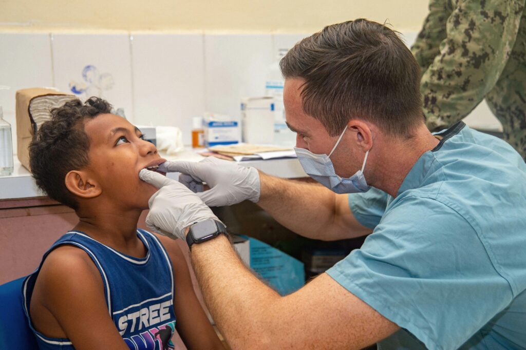
Pediatric Dentistry Guide · For Parents · Williamsburg Your dentist said “baby root canal” and your stomach dropped. Here’s what a pulpotomy actually is, why it’s far less dramatic than it sounds, and what to expect step by step. Does My Child Really Need a Pediatric Root Canal? A Parent’s Guide WHY WE SAVE BABY TEETH WHAT HAPPENS DURING THE VISIT Written by Toothology Editorial Team | Reviewed by Dr. Ayala Lieberman, DDS June 2026 Your child has been up through the night with tooth pain. Or your pediatric dentist just mentioned the words “baby root canal” at a routine checkup — and your stomach dropped a little. Both reactions are completely understandable. But a pediatric root canal sounds more alarming than it usually is. The procedure most commonly recommended for young children is actually a pulpotomy — a gentler, more limited treatment than the adult version most parents picture. When a primary tooth has decay that has reached the pulp (the soft tissue inside the tooth containing nerves and blood vessels), a pulpotomy removes the infected portion of that tissue rather than the entire pulp structure. The goal is to save the tooth, relieve the pain, and protect the underlying permanent tooth that is still developing beneath it. WHAT IS A PULPOTOMY? A pulpotomy removes only the infected pulp in the crown (top portion) of the baby tooth — not the roots. This is why it’s usually quicker and less involved than the adult root canal most parents are imagining. It’s one of the most common pediatric dental procedures. Why We Must Save a Baby Tooth with a Pulpotomy It’s a common question from parents: if the tooth will fall out anyway, why not just pull it? The short answer is that back baby teeth do important work for years — often until ages 10–12 for back molars. Pulling them early creates real problems. Chewing & Nutrition Primary molars handle most chewing in childhood. Losing them early can make eating harder and significantly limit food choices during key developmental years. Speech Development Teeth help children form certain sounds. Early tooth loss can affect speech clarity while speech patterns are still developing and forming. Space for Adult Teeth Primary molars hold the space for the adult tooth underneath. If removed too early, nearby teeth drift — often leading to crowding and orthodontic treatment later. Bone & Tissue Preservation Keeping the tooth helps maintain the bone and gum support in that area until the permanent tooth is ready to erupt naturally. Pulpotomy vs. Extraction vs. Adult Root Canal Option What Happens Goal & Typical Follow-Up Pulpotomy Baby root canal Infected pulp in the crown of the tooth is removed; a medicated mineral-based filling is placed to protect the remaining healthy tissue Save the baby tooth and restore function; crown typically placed the same appointment or soon after; tooth held in place until it falls out naturally Extraction Tooth removal The primary tooth is removed under local anaesthetic; straightforward when the tooth cannot be restored or infection is too advanced Eliminate infection quickly; a space maintainer is often needed to hold the gap until the permanent tooth erupts — without one, nearby teeth can drift and cause crowding Adult Root Canal For permanent teeth Full pulp removed — including the root canals; canals are shaped, cleaned, and sealed; a different and more involved process than a pulpotomy Save a permanent tooth for life; typically followed by a crown; not the same procedure as a pulpotomy — and generally longer and more complex A pulpotomy only removes infected tissue from the top (crown of the tooth — not the root canals. This is why it’s usually quicker and less involved than the adult root canal most parents picture. WHEN EXTRACTION MAY BE RECOMMENDED INSTEAD A pulpotomy isn’t always the best option. If the tooth can’t be restored — because decay is too extensive, infection has affected surrounding bone, or there isn’t enough healthy tooth left to rebuild — extraction may be recommended. Your dentist should explain why they’re recommending extraction vs. saving the tooth. It’s reasonable to seek a second opinion for major decisions. Signs Your Child Has a Dental Infection Deep in the Tooth Tooth decay that is still in the outer layers of a tooth rarely causes significant pain. When decay reaches the pulp, the symptoms change — and they tend to be hard to miss. Toothache that wakes your child at night or is persistent rather than occasional Sharp or lingering sensitivity to hot, cold, or sweet foods and drinks Spontaneous pain — throbbing or sharp pain without any food or drink trigger A small pimple or bump on the gum near a tooth — this may be a dental abscess draining Swelling of the gum or face near the affected tooth A persistent bad taste in the mouth that your child mentions Refusal to chew on one side of the mouth Visible darkening of a tooth that was previously a normal colour Fever accompanying dental symptoms What Is a Dental Abscess and Why Does It Matter? A dental abscess is a pocket of infection that forms when decay reaches the tooth’s nerve and spreads into nearby tissue or bone. In children, it may show up as a small pimple-like bump on the gum (a draining fistula). Even if that bump seems to reduce pain — it doesn’t mean the infection is gone. It means it’s still active and draining. Dental infections don’t resolve on their own. Without treatment, they can spread to nearby teeth and bone and may affect the developing adult tooth underneath. When to Call 911 or Go to the ER Instead Facial or neck swelling that is spreading , especially toward the jaw, eye, or throat Difficulty breathing or swallowing — this is a medical emergency, not a dental emergency High fever alongside significant facial swelling Swelling that changes rapidly over hours rather than days Step-by-Step:
Should You Get a Second Opinion on Your Dental Treatment Plan?

Patient Guidance · Treatment Planning When a treatment plan feels bigger than expected, a second opinion is not distrust — it is the most rational way to approach a decision that is significant in cost, impact, or permanence. Should You Get a Second Opinion on Your Dental Treatment Plan? Written by Toothology Editorial Team | Reviewed by Dr. Ayala Lieberman, DDS May 2026 You walk out of a dental appointment holding a treatment plan that lists procedures you were not expecting — several crowns, a root canal, possible extractions, or a full-mouth reconstruction that comes with a price tag that makes you hesitate at the door. You trust your dentist well enough. But something about the conversation felt rushed, or the scale of the recommended work surprised you, or you simply want to understand your options before committing to something this significant. Seeking a dental second opinion in that situation is not an act of distrust. It is a reasonable, patient-centred step that any responsible dental professional should welcome. Dental Emergency? If you are dealing with a dental emergency — active infection, significant swelling, severe pain — please seek evaluation promptly rather than waiting for a second opinion to be arranged. Why High-Cost Dental Work Often Warrants a Second Look There is nothing inherently suspicious about receiving a large treatment plan from a dentist. Dental disease is progressive, and it is genuinely common for patients who have deferred care for a few years to discover multiple problems at once. Situations Where a Second Opinion Is Particularly Worth Pursuing You have been quoted for five or more crowns, implants, or major restorations at once The consultation felt rushed and you left without a clear understanding of what is wrong Created with Sketch Beta. You have been recommended for full-mouth rehabilitation, All-on-X, or a significant bite reconstruction You have been told to extract a tooth you were not expecting to lose — without a clear explanation The treatment plan includes procedures you have not been given alternatives for Recommendations have changed significantly between dentists without clear explanation You feel pressure to commit quickly or offered heavy discounts for same-day decisions A large root canal or surgical procedure is recommended and you want to understand the basis Urgency Changes the Calculation Not every dental situation can wait. Swelling, spreading infection, significant pain, or any sign of a dental abscess are situations where prompt evaluation and treatment are clinically appropriate. If your dentist has expressed any urgency around a treatment recommendation, take that seriously and discuss the timeline openly. The Four Things a Good Second Opinion Should Confirm 1 The Diagnosis Is the clinical finding — the decay, fracture, bone loss — accurately identified and clearly documented? 2 The Alternatives Is the recommended treatment the only reasonable option, or are there alternatives with different risk/benefit profiles? 3 The Urgency Does each component need to be addressed immediately, or is phased treatment over time a viable option? 4 The Risks of Deferral If you wait, what realistically happens? Understanding the cost of inaction is as important as the cost of action. Treatment transparency — understanding the evidence behind each recommendation — is something every patient is entitled to, and something a thorough second opinion should provide. Using Advanced 3D Technology for Accurate Diagnosis The quality of a second opinion is only as good as the quality of the diagnostic information underlying it. A quick visual exam alone offers far less clarity than a review that includes current radiographs, a thorough clinical assessment, and — when appropriate — 3D imaging. What a High-Quality Second-Opinion Exam Typically Includes Review of existing records: X-rays, treatment plan, itemised estimate, and any clinical notes from your first provider Updated or supplemental radiographs: If your existing X-rays are recent and of good quality, they may be sufficient Comprehensive clinical examination: A full assessment of teeth, gums, bite, and any symptomatic areas — not just the teeth already identified Periodontal charting: For any plan involving gum disease treatment or implants, a complete picture of bone levels and pocket depths 3D imaging (CBCT) when clinically indicated: For complex implant planning, suspected root fractures, bone volume assessment, or nerve/sinus proximity A clear, written explanation of findings: What the clinician found, what they recommend, what the alternatives are, and the risks of each path When 3D Imaging Makes a Material Difference Implant Planning Assessing bone volume, quality, and the precise location of the inferior alveolar nerve or maxillary sinus before placing a titanium post is significantly more accurate in three dimensions. Suspected Root Fractures Vertical root fractures are frequently missed or inconclusive on standard radiographs. CBCT can reveal fracture patterns that change whether a tooth is restorable or should be extracted. Root Canal vs. Extraction The three-dimensional anatomy of root canals, periapical pathology, and surrounding bone is more accurately assessed in 3D — particularly for multi-rooted posterior teeth. Bone Level Assessment The extent and pattern of bone loss in periodontal disease is more completely characterised in three dimensions than on flat radiographs. Not Every Patient Needs 3D Imaging The decision to use CBCT should be clinically justified by the complexity of the case and the diagnostic question that needs answering — the benefit should outweigh the additional radiation exposure, which while low relative to medical CT is not zero. Common Procedures Where a Second Opinion Saves Money While a second opinion is not a guarantee of a lower-cost alternative — the original plan may well be confirmed as the right approach — there are specific clinical situations where the treatment plan can reasonably vary between practitioners, and where understanding the full option set is genuinely valuable before committing. Situation Option A Option B What to Ask Single missing tooth Dental Implant Fixed, preserves bone, most closely mimics natural tooth Bridge Implant-supported or traditional bridge — no surgery, lower initial cost What are the long-term bone implications of each? What is the risk to adjacent teeth? Tooth with deep infection / structural damage Root Canal Preserves the natural tooth root, allows crown placement Extraction +